


Hepatitis E Virus (HEV) is an infection known worldwide for its asymptomatic and self-limited course in most cases. Some cases progressing to chronicity have been described in immunosuppressed patients, especially in recipients of solid organ transplants. We evaluated laboratory parameters of HEV infection (HEV RNA, anti-HEV IgM and anti-HEV IgG) through enzyme-linked immunosorbent assay (Elisa), confirmed by immunoblotting, in a cohort of 294 patients who received liver transplants at the HCFMUSP (Hospital das Clínicas da Faculdade de Medicina da Universidade de São Paulo). Laboratory and demographic data were collected from the entirety of the transplanted population. Hepatic biopsies of 122 patients transplanted due liver failure secondary to hepatitis C (HCV), with or without serological or molecular markers of HEV, were analyzed according to METAVIR score. Out of 24 (8.2%) patients tested positive for anti-HEV IgG, six (2%) were positive for anti-HEV IgM and 17 (5.8%) for HEV RNA. Of the patients transplanted because of HCV infection, 95 (77.8%) had received treatment including ribavirin for at least six months before blood sample collection. Among patients transplanted due to HCV cirrhosis who tested positive for anti-HEV IgG, only three (37.5%) showed fibrosis beyond stage 2, while five (41.7%) of the HEV RNA-positive patients had liver fibrosis beyond stage 2. Overall, the prevalence of HEV in the post-hepatic transplant scenario appears to be low, and, at least histologically, seemingly not harmful. We conclude that, although some studies reported a risk of HEV chronification, patients who had their livers transplanted due to HCV and showed serological or molecular markers of HEV did not have higher levels of fibrosis compared to patients who showed no indications of HEV infection at the time of the analysis.
Hepatitis E virus (HEV) infection is a public health problem in developing countries, presenting as both sporadic and epidemic cases. Data from the World Health Organization (WHO) show that approximately 3.7 million people worldwide are infected with the virus and mortality can reach about 70,000 people per year.1 At least five different genotypes have been described. However, only four are able to infect human beings. Genotype 3 has been frequently associated with infections in animals, while genotypes 1 and 2 primarily infect human beings.2 Most patients infected with HEV live in endemic regions, underdeveloped countries where the main route of transmission is fecal-oral. However, an increasing number of infections, especially genotype 3, has been reported in developed countries, with the main route of dissemination through consumption of raw or undercooked pork.2,3 HEV infection usually induces acute, oligosymptomatic and self-limiting hepatitis in immunocompetent subjects. Although recent cases of evolution to chronicity caused by HEV have been identified in immunosuppressed subjects such as patients with HIV, malignant hematologic diseases and transplanted solid organs.4 In liver transplant patients, cases of acute hepatitis of unknown etiology with persistently elevated levels of transaminases should be investigated using molecular tests for HEV RNA since anti-HEV IgG seroconversion can be delayed or never even occur.5,6
On the other hand, hepatitis C virus (HCV) remains a public health problem with approximately 3% of the world population being infected.7 The incidence and consequence of HEV superinfection among HCV patients is unknown and could be a contributing factor for liver decompensation increasing morbidity and mortality among patients. The objective of this study was to evaluate the prevalence of HEV infection among liver transplant patients of several etiologies in our country, using serological or molecular markers of HEV infection, as well as to evaluate the role of HEV infection under the histological damage of patients with of hepatitis C virus infection after liver transplantation followed at the outpatient clinic of Hospital das Clínicas da Universidade de São Paulo (HCFMUSP), Brazil.
Patients and methodsStudy subjectsA descriptive cross-sectional study was conducted to determine the prevalence of serological and molecular markers in all liver transplant patients of any etiology. A comparative analysis of liver biopsies of patients transplanted due to HCV with or without serological and/or molecular markers of HEV was conducted. A total of 294 liver transplant patients who had been transplanted for more than six months were enrolled between January 2008 and January 2013. All were regularly followed at HCFMUSP outpatient clinic. The patients of either sex, over 18 years old were included. Transplantation had been performed due to acute or chronic liver disease of any etiology, including liver cancer. All had compensated liver disease. Serological and molecular tests performed were anti-HEV IgM, anti-HEV IgG and HEV-RNA. Levels of AST, ALT, and GGT were investigated by the automated kinetic method used in the hospital laboratory routine, with normal values of ALT up to 41 IU/L for male and 30 IU/L for female, AST up to 37 IU/L for male and 31 IU/L for female, and GGT up to 60 IU/L in male and 36 IU/L in female. A comparative study was performed between liver biopsies of 122 patients transplanted due to chronic HCV infection, whether or not they had serological or molecular markers of HEV infection, according to the METAVIR8 classification. Due to the small number of positive samples for anti-HEV IgM, anti-HEV IgG and HEV RNA were used for a statistical analyses, and the histological indices of inflammatory activity and fibrosis from METAVIR classification were pooled into the following categories: stage 1, degrees of 0 and 1; stage 2, activity degrees 2, 3 and 4. Patients could have been treated for hepatitis C with peginterferon with or without ribavirin for different periods, depending on HCV genotype and viral kinetics.
The study was approved by the HCFMUSP Research Ethics Committee, and conducted in accordance with the provisions of the Declaration of Helsinki.
Serological tests for HEV infectionELISA methodology (RecomWell HEV IgM/IgG from Mikrogen®) was used for antibody screening. Indeterminate results or isolate positive IgM were confirmed by immunoblotting (RecomLine IgM/IgG from Mikrogen®). One-step real-time PCR methodology with primers and probes that amplify an ORF3 fragment of the viral genome was used for HEV RNA survey.
Histological evaluationLiver biopsies selected for the study were at least 15 mm long and had at least 10 porta hepatis. In addition, to avoid variability among observers, all samples were reviewed by an experienced pathologist, without knowledge of the clinical condition and the laboratory tests of the patients. Histological Activity Index (IAI) was classified according to the METAVIR8 score since it is widely used worldwide.
Statistical analysisA univariate analysis was performed to assess the clinical and laboratory variables of all liver transplant recipients. A multivariate analysis was performed to assess the relationship between serologic and molecular markers with disease etiology and degrees of inflammation and fibrosis. Chi-square test with Yates correction was used for comparisons between groups. A p<0.05 was considered statistically significant.
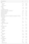
ResultsOf 294 transplanted patients, 179 (60.9%) were male, majority in the age range 40–59 years (n=117, 39.8%). Cirrhosis due to viral causes (HCV, HBV and HBV/HCV co-infection) was the main indication for liver transplantation with 162 (55.1%) cases, followed by cirrhosis due to cryptogenic, autoimmune and vascular diseases in 53 (18%) patients.
Anti-HEV IgG serum marker was positive in 24 (8.2%) cases, while anti-HEV IgM was positive in six (2%), with only one patient showing simultaneous positivity for both markers. HEV RNA was detected in 17 (5.8%) patients and in only one of them anti-HEV IgG was also detected.
AST levels were abnormal in 25.2% (74) of the cases, ALT in 31.6% (93), and GGT in 58.5% (172) (Table 1).
General characteristic of the liver transplant patients submitted to HEV investigation.
HVB, Hepatitis B virus; HCV, Hepatitis C virus; NASH, Non-Alcoholic Steatohepatitis; ASH, Alcoholic Steatohepatitis; PBC, Primary Biliary Cholangitis; AIH, Autoimmune Hepatitis; HEV RNA, Hepatitis E virus ribonucleic acid (positive or negative); ALT, Alanine Aminotransferase (normal range: up to 41 in males, up to 30 in females); AST, Aspartate Aminotransferase (normal range: up to 37 in males, up to 31 in females); GGT, Gamma Glutamyl Transferase.
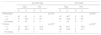
Among the main etiologies leading to transplantation, chronic viral infections presented the highest positivity rates of anti-HEV IgG and HEV-RNA, with 15 (62.5%) and with 13 (76.5%) cases, respectively (Table 2).
Evaluation of serological (anti-HEV IgG) and molecular (HEV RNA) markers according to the liver disease etiology leading to transplantation.
HVB, Hepatitis B virus; HCV, Hepatitis C virus; NASH, Non-Alcoholic Steatohepatitis; ASH, Alcoholic Steatohepatitis; PBC, Primary Biliary Cholangitis; AIH, Autoimmune Hepatitis.
Of 294 patients included, 122 (41,5%) had chronic hepatitis C as their indication for transplantation; of those, 95 (32.3%) had been treated for HCV before the time of blood collection, five (1.7%) were untreated, and 22 (7.5%) had no treatment information or had discontinued treatment follow-up. The remaining 172 (58.5%) patients did not have chronic hepatitis C as the cause of the transplant.
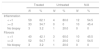
Regarding the histological findings, four (50%) of the anti-HEV IgG positive patients presented inflammation stages lower than or equal to 1, and four (50%) presented inflammation stages higher than 2. Of the HEV RNA-positive patients, six (50%) showed inflammation stages lower than or equal to 1, while six (50%) patients had stages higher than 2. For these two markers, there was no significant difference (p > 0.05) when compared with the negative results in both stages.
Regarding fibrosis, five (62.5%) of the anti-HEV IgG-positive patients presented stages lower than or equal to 1, whereas three (37.5%) showed stages higher than 2. In relation to the HEV positive RNA patients, seven (58.3%) had fibrosis stages lower than or equal to 1, while five (41.7%) presented stages higher than 2. For these two markers, there was no significant difference (p > 0.05) when compared with the negative results in both stages (Table 3).
Correlation between histological findings of patients with HCV and positive serological or molecular markers for HEV infection according to METAVIR score.
HEV RNA, Hepatitis E virus ribonucleic Acid; Neg, Negative; Pos, Positive; Val, Absolute value; Prop, Proportion.
Of 95 patients treated for HCV, 59 (62.1%) had inflammation stages lower than or equal to 1 and 33 (34.7%) had inflammation stages higher than 2. Among patients not treated for HCV, four (80%) showed degrees of inflammation lower than or equal to 1 and none of them presented degrees of inflammation higher than or equal to 2 (p > 0.05). In relation to grade of fibrosis, 40 (42.1%) of the treated patients presented fibrosis less than or equal to stage 1, while 52 (54.7%) had fibrosis beyond stage 2. In three (60%) of the untreated patients, fibrosis levels were lower than or equal to 1, while only one (20%) had fibrosis beyond stage 2 (p > 0.05) following re-categorization of the METAVIR classification (Table 4).
Correlation between histological findings of patients submitted or not to HCV treatment according to METAVIR score.
N/A, No Answer; Neg, Negative; Pos, Positive; Val, Absolute value; Prop, Proportion.
Hepatitis E is a globally distributed disease, and is the leading cause of acute non-A, non-B viral hepatitis worldwide. In our study, the prevalence of anti-HEV IgG positivity in liver transplant patients was 8.2%, while that of anti-HEV IgM was 2%. However, in most of studies evaluating the role of HEV chronicity in liver transplant patients anti-HEV IgG found a somewhat lower frequency1 than in the present study, about 1–3%. It is important to consider the significant social-cultural differences between the countries studied.1 A Dutch study evaluated 285 patients undergoing liver transplantation who were investigated for anti-HEV, anti-HEV IgG, and HEV RNA;9 96% of these individuals were negative for all markers, and only nine (3.1%) presented anti-HEV IgG positivity, demonstrating that there was no evidence of HEV infection in the post-transplant period in this group. French studies also investigated the prevalence of HEV in liver transplant patients between the years 2005 to 2012, having found anti-HEV IgG in 7.7% of patients after an average follow-up of 33 months, a very similar number to what was found in our case-by-case analysis.10 In a Spanish study conducted by Buti et al. in 2010, the prevalence of HEV was investigated in 82 liver transplant patients through anti-HEV IgG in those with hepatic enzyme elevations. In patients who were anti-HEV IgG-positive, anti-HEV IgM and HEV RNA were also tested. Only three (3.65%) patients were positive for anti-HEV IgG, having no evidence of persistent infection in these cases.11 Pischke et al.4 showed, in a population of 226 liver transplant patients, 4.5% positivity for anti-HEV IgG, 156 of which showed no evidence of hepatitis in the graft. Thus, the prevalence of present or past HEV infection in liver-transplanted patients appears to be low in several case-by-case analyses. Heterogeneity may in part be justified by the difference in sensitivity of the serological kits used in the various studies. U.S. studies conducted by Fontana et al.12 showed the role of the hepatitis E virus among 681 American adults with acute liver failure, testing for anti-HEV IgM and anti-HEV IgG. Subjects positive for anti-HEV IgM were also tested for HEV RNA. At admission, 294 (35.2%) of patients with acute liver failure were anti-HEV IgG positive. Only three patients showed anti-HEV IgM positive, but all of them were negative for HEV RNA. Thus, the authors concluded that acute hepatic failure by HEV is rare in American adults and should not be related to causes of indeterminate hepatitis. In our series, the positivity for HEV RNA was 5.8%, very similar to what was found in the published literature. However, the incidence of HEV infection based on the detection of HEV RNA can range from 0.9% to 3.5%, according to Kamar et al.13
In our analysis, AST levels were abnormal in 25.2% of cases, ALT in 31.6% and GGT in 58.5% in all transplanted patients. ALT levels were altered in 41.7% of patients who were anti-HEV IgG-positive, compared to 31.1% of those with normal levels, while 37.5% had changes in AST levels compared to 24.4% who presented levels within normal range. We also found 10 (58.8%) patients with abnormal ALT levels with positive HEV RNA, while in 47.1% AST levels were normal. A Chinese study evaluating 5023 blood donors with elevated ALT levels showed a 33.3% prevalence of anti-HEV IgG, which was significantly higher than the 24.9% found in 4046 donors who had normal ALT levels (p < 0.05). The prevalence of anti-HEV IgM was similar among donors with normal levels of ALT, 60 of 4026 (1.48%) and donors with high ALT, 71 of 5023 (1.41%) [p > 0.05].14 HEV RNA was present in only six donors that had elevated levels of ALT.14 This might translate into lower inflammatory activity even in those who were positive for HEV infection.
Another widely discussed aspect is the natural history of the progression to HEV chronicity, which is not yet fully understood, making it difficult to evaluate the mechanisms responsible for this occasional progression. When assessed for fibrosis by METAVIR classification, the numbers were similar, showing that 37.5% of patients with anti-HEV IgG positive had fibrosis levels higher than 2, while 41.7% of patients with HEV positive RNA had fibrosis levels higher than 2 (p > 0.05) when compared with patients negative for these HEV infection markers; nevertheless, the difference was not statistically significant. However, one of the first series of cases showing chronic HEV infection in transplanted subjects included 14 solid-organ receptors. Overall, eight patients developed persistent infection, including three (21.4%) patients who underwent liver transplantation.15 Subsequently, a number of other specific studies have been published reporting the role of chronic HEV infection in organ transplant recipients, including liver transplanted patients, with similar results, namely a low absolute frequency of 1–3% in most studies, despite differences in epidemiology, climate, and even viral strains among the countries studied. In a classic study with 226 liver transplant patients, anti-HEV IgG was diagnosed in 10 (4.4%) cases, with HEV RNA being positive in three (1.3%) of them. Of these three patients, two developed persistent viremia, with only one progressing to advanced fibrosis 22 months after liver transplantation.16
On the other hand, it is believed that HEV superinfection among patients with chronic liver disease could cause liver decompensation leading to increased morbidity and mortality.7 In our series, of 122 transplanted patients due to HCV infection, 95 (32.3%) were treated with regimens that included ribavirin before the collection of our samples. Of the 122 treated patients, 33 (34.7%) had levels of inflammatory activity higher than 2 and 52 (54.7%), had fibrosis levels higher than 2, according to the re-categorization of the METAVIR classification; however, these data were not statistically significant when compared to patients who had not been treated for HCV.7 Thus, in our analysis, patients co-infected with HCV and HEV did not present levels of inflammatory activity or fibrosis significantly higher than patients infected with HCV alone. In addition, patients treated for HCV with ribavirin also did not present significantly higher levels of inflammatory activity and fibrosis when compared to those not treated with ribavirin, showing that in our setting HEV infection appeared not to be responsible for increased inflammatory damage or fibrosis among HCV liver transplant patients co-infected with HEV at the time of analysis.
ConclusionAlthough the diagnosis of hepatitis E should be considered in the population of liver transplant patients who develop hepatitis of unknown etiology, this study showed that the prevalence of HEV circulation in our country is low when compared to European countries, based on a survey of anti-HEV IgM, anti-HEV IgG and HEV RNA. Liver transplant recipients who tested positive for these markers did not have higher levels of AST, ALT or GGT when compared to patients with negative serological or molecular markers for HEV infection. Among liver transplant patients with chronic hepatitis C who presented positive serological or molecular markers for HEV infection, there was no greater histological damage, whether in indexes of inflammatory activity or stages of fibrosis, when compared to those who had negative markers for HEV infection, showing a lower inflammatory activity.
Therefore, in our setting, HEV and HCV co-infection does not appear to have an impact on worsening of the histological progression of liver disease in the post-transplantation scenario. However, prospective studies with more robust patient samples and longer follow-up are necessary.
Financial disclosureThis research did not receive any specific grant from funding agencies in the public, commercial, or not-for-profit sectors.
