To characterize methicillin-resistant Staphylococcus aureus isolates from an intensive care unit of a tertiary-care teaching hospital, between 2005 and 2010. A total of 45 isolates were recovered from patients admitted to the intensive care unit in the study period. Resistance rates higher than 80% were found for clindamycin (100%), erythromycin (100%), levofloxacin (100%), azithromycin (97.7%), rifampin (88.8%), and gentamycin (86.6%). The SCCmec typing revealed that the isolates harbored the types III (66.7%), II (17.8%), IV (4.4%), and I (2.2%). Four (8.9%) isolates carried non-typeable cassettes. Most (66.7%) of the isolates were related to the Brazilian endemic clone from CC8/SCCmec III, which was prevalent (89.3%) between 2005 and 2007, while the USA100/CC5/SCCmec II lineage emerged in 2007 and was more frequent in the last few years. The study showed high rates of antimicrobial resistance among methicillin-resistant S. aureus isolates and the replacement of Brazilian clone, a well-established hospital lineage, by the USA100 in the late 2000s, at the intensive care unit under study.
Staphylococcus aureus is one of the main causes of healthcare-associated infections.1 Methicillin-resistant S. aureus (MRSA) is a growing problem worldwide and is associated with significant morbidity, mortality and increased costs of treatments.2,3 The majority of MRSA isolates are found in intensive care units (ICUs).4 In Brazil, these rates have even reached 70%.5 Methicillin resistance is located in a staphylococcal cassette chromosome (SCCmec) and the most frequent types are II, III and IV.2
Recent studies have described the emergency of MRSA lineages in hospitals worldwide.6,7 In Brazil, although isolates related to the Brazilian endemic clone (BEC)/SCCmec III/CC8 have caused the majority of hospital-acquired staphylococcal infections in the past,8 during the last decade an increased occurrence of nosocomial infections due to isolates carrying the SCCmec IV and II have been described.9–11 The characterization of MRSA isolates from patients in ICUs has already been described by various authors.9,11 However, these studies only analyzed a few ICU isolates within a larger hospital collection, without highlighting the characterization of these isolates. This study investigated the phenotypic and molecular characteristics of a collection of MRSA clinical isolates from patients admitted to a Brazilian ICU over a six-year period.
The study was conducted at a tertiary-care teaching hospital affiliated to the Federal University of Juiz de Fora, Minas Gerais, Brazil. This is a 146-bed, six of them being ICU. The MRSA isolates were obtained from patients admitted to the ICU consecutively, between 2005 and 2010, from different sources, such as tracheal secretion (53.3%), blood (20%), catheter tip (13.4%), and others (13.3%). Bacterial identification and susceptibility to methicillin were determined at the hospital laboratory by the classical identification tests12 and the cefoxitin (Oxoid, Basingstoke, UK) disk diffusion test,13 respectively. Minimum inhibitory concentration (MIC) was assessed for 11 drugs as recommended by the CLSI.13 Bacterial DNA was extracted as previously described14 and the determination of the SCCmec types was performed according to Milheiriço et al.15 All MRSA isolates were typed by PFGE8 and the clonality was determined according to Van Belkum et al.16 criteria using previously characterized control strains, such as: USA100/SCCmec II, USA400 and USA800/SCCmec IV9 and BEC/SCCmec III.8 One isolate representative of each PFGE profile was chosen for characterization of the clonal complex (CC).17 Statistical comparisons were performed by analysis of contingency tables using Fisher's exact test; level of significance was established at 5% (p<0.05).
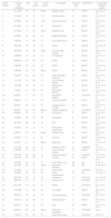
Out of 76 S. aureus recovered from patients admitted to the ICU, between January/2005 and November/2010, 45 (59.2%) MRSA isolates recovered from 45 patients were evaluated. Except for vancomycin and linezolid, whose MIC90 was 2.0μg/mL, high rates of resistance were found for seven of the 11 antimicrobials tested (Table 1). Among the 45 MRSA isolates, 30 (66.7%) harbored the SCCmec III, 8 (17.8%) the type II, 2 (4.4%) the type IV and 1 (2.2%) the type I. Four (8.9%) MRSA isolates were non typeable (NT). S. aureus isolates carrying SCCmec III were related to the BEC/clonal complex (CC) 8 (Table 2). All the eight isolates that carried the SCCmec II were related to the USA100/CC5 lineage. Two isolates carrying the SCCmec IV belonged to the lineages USA400/CC1 and USA800/CC5. The SCCmec I isolate was associated to USA500/CC5 and the other four MRSA isolates did not belong to any clonality previously described (Table 2).
Antimicrobial resistance of 45 MRSA isolates recovered from patients of an ICU at a Minas Gerais teaching hospital, between 2005 and 2010.
| Antimicrobial agent | Minimal Inhibitory Concentration (μg/mL) | No (%) of resistant isolates | ||
|---|---|---|---|---|
| MIC50 | MIC90 | Range | ||
| Azithromycin | >1024.0 | >1024.0 | 0.5–>1024.0 | 44 (97.7) |
| Chloramphenicol | 32.0 | 64.0 | 4.0–128.0 | 29 (64.4) |
| Clindamycin | >1024.0 | >1024.0 | 512.0–>1024.0 | 45 (100) |
| Erythromycin | 512.0 | 512.0 | 256.0–512.0 | 45 (100) |
| Gentamicin | 128.0 | 1024.0 | 0.125–>1024.0 | 39 (86.6) |
| Levofloxacin | 4.0 | 16.0 | 2.0–32.0 | 45 (100) |
| Linezolid | 2.0 | 2.0 | 1.0–2.0 | 0 (0) |
| Rifampin | 2.0 | 256.0 | 0.0625–>1024.0 | 40 (88.8) |
| Tetracycline | 32.0 | 64.0 | 0.0625–128.0 | 31 (68.8) |
| Trimethoprim/sulfamethoxazole | 32.0/608.0 | 128.0/2432.0 | 0.0625/2.3–1024.0/19,456.0 | 32 (71.1) |
| Vancomycin | 1.0 | 2.0 | 0.5–2.0 | 0 (0) |
MIC50, minimal inhibitory concentration that inhibits 50% of bacterial population; MIC90, minimal inhibitory concentration that inhibits 90% of bacterial population.
Characteristics of 45 MRSA isolated from patients admitted to an ICU of a Minas Gerais teaching hospital, between 2005 and 2010.
| Isolate number | Isolation date (mm/dd/yy) | Bed | Age (years) | Clinical source | Comorbidity | SCCmec type | Clonalitya/CC | Antimicrobial resistance profile |
|---|---|---|---|---|---|---|---|---|
| 1 | 01/11/05 | 04 | 77 | Blood | Bladder cancer | III | BEC/8 | A C E G L R S T |
| 2 | 01/18/05 | 01 | 79 | TS | Stomach cancer | III | BEC/8 | A C E G L R S T |
| 3 | 03/08/05 | 03 | 35 | Urine | Respiratory failure | III | BEC/8 | A H C E G L R S T |
| 4 | 05/17/05 | 03 | 78 | TS | Pneumonia | III | BEC/8 | A H C E G L R S T |
| 5 | 07/18/05 | 01 | 69 | Blood | Bladder cancer | III | BEC/8 | A H C E G L R S T |
| 6 | 07/24/05 | 02 | 40 | TS | Respiratory failure | III | BEC/8 | A C E G L R S T |
| 7 | 07/25/05 | 01 | 48 | TS | Incarcerated hernia | III | BEC/8 | A C E G L R S T |
| 8 | 08/31/05 | 04 | 25 | PL | Ascitis | NT | ND/ND | A C E G L R S T |
| 9 | 09/08/05 | 03 | 69 | Blood | Coronary artery disease | III | BEC/8 | A H C E G L R S T |
| 10 | 09/06/06 | 05 | 75 | CT | Fahr disease | III | BEC/8 | A C E G L R S T |
| 11 | 09/23/06 | 02 | 66 | TS | COPD | III | BEC/8 | A C E G L R S T |
| 12 | 10/09/06 | 02 | 60 | TS | COPD | III | BEC/8 | A H C E G L R S T |
| 13 | 12/06/06 | 03 | 81 | Blood | COPD | III | BEC/8 | A C E G L R S T |
| 14 | 12/09/06 | 04 | 23 | TS | Acute intermittent porphyria | III | BEC/8 | A H C E G L R S T |
| 15 | 12/11/06 | 05 | 69 | TS | Chronic kidney disease | III | BEC/8 | A H C E G L R S T |
| 16 | 12/18/06 | 03 | 53 | CT | Rheumatoid arthritis | III | BEC/8 | A H C E G L R S T |
| 17 | 01/30/07 | 01 | 53 | TS | Rheumatoid arthritis | III | BEC/8 | A H C E G L R S T |
| 18 | 02/04/07 | 01 | 56 | TS | Neurogenic bladder | II | USA100/5 | A H C E G L R |
| 19 | 02/28/07 | 01 | 41 | PL | Acute pancreatitis | III | BEC/8 | A H C E G L R S T |
| 20 | 03/25/07 | 02 | 76 | TS | Cerebrovascular accident | III | BEC/8 | A H C E G L R S T |
| 21 | 04/02/07 | 04 | 54 | Blood | Hodgkin lymphoma | II | USA100/5 | A C E G L R |
| 22 | 06/25/07 | 03 | 61 | CT | Hemothorax | III | BEC/8 | A H C E G L R S T |
| 23 | 07/08/07 | 04 | 42 | SS | Pancreatitis | III | BEC/8 | A H C E G L R S T |
| 24 | 09/10/07 | 06 | 35 | TS | Aids | III | BEC/8 | A H C E G L R S T |
| 25 | 09/18/07 | 03 | 74 | TS | Extrapontine myelinolysis | III | BEC/8 | A H C E G L R S T |
| 26 | 12/04/07 | 04 | 73 | Blood | Bladder cancer | III | BEC/8 | A H C E G L R S T |
| 27 | 12/24/07 | 01 | 51 | PL | Cirrhosis | III | BEC/8 | A H C E G L R S T |
| 28 | 12/31/07 | 01 | 73 | Blood | Malnutrition | III | BEC/8 | A C E G L R S T |
| 29 | 01/14/08 | 02 | 23 | CT | Diabetes mellitus | IV | USA800/5 | C E G L T |
| 30 | 01/30/08 | 03 | 65 | TS | Chronic kidney disease | III | BEC/8 | A H C E L R S T |
| 31 | 02/03/08 | 02 | 84 | TS | Respiratory failure | IV | USA400/1 | A H C E G L |
| 32 | 02/07/08 | 06 | 59 | CT | Cirrhosis | III | BEC/8 | A H C E G L R S T |
| 33 | 03/16/08 | 03 | 81 | CT | Acute kidney injury | NT | ND | A C E L S |
| 34 | 08/16/08 | 06 | 61 | Blood | Myocardial infarction | NT | ND | A C E G L R |
| 35 | 10/13/08 | 01 | 65 | SS | Appendicitis | I | USA500/5 | A C E G L |
| 36 | 07/18/09 | 03 | 42 | TS | Renal transplantation | NT | ND | A H C E L R |
| 37 | 08/03/09 | 05 | 19 | TS | Pneumonia | II | USA100/5 | A H C E L R |
| 38 | 08/09/09 | 01 | 78 | TS | Breast cancer | II | USA100/5 | A H C E G L R |
| 39 | 08/17/09 | 06 | 79 | TS | COPD | II | USA100/5 | A H C E G L R |
| 40 | 12/14/09 | 01 | 83 | TS | Cholangitis | III | BEC/8 | A H C E G L R S T |
| 41 | 02/06/10 | 02 | 44 | TS | Acute kidney injury | II | USA100/5 | A H C E G L R |
| 42 | 02/21/10 | 04 | 98 | TS | Aplastic anemia | III | BEC/8 | A H C E G L R S |
| 43 | 04/18/10 | 06 | 35 | TS | Aids | III | BEC/8 | A H C E G L R S T |
| 44 | 08/25/10 | 06 | 59 | Blood | Necrosis in amputation | II | USA100/5 | A C E L |
| 45 | 11/30/10 | 04 | 62 | TS | Acute kidney injury | II | USA100/5 | A C E L R |
CT, cateter tip; PL, peritoneal liquid; SS, surgical site; TS, tracheal secretion; COPD, chronic obstructive pulmonar; CC, clonal complex; NT, non typeable; ND, not determined; A, Azithromycin; C, Clindamycin; E, Erythromycin; G, Gentamicin; H, Chloramphenicol; L, Levofloxacin; R, Rifampin; S, Trimethoprim/sulfamethoxazole; T, Tetracycline.
Brazilian studies have evaluated the epidemiology of MRSA and the results indicate that several lineages initially restricted to other continents are emerging in Brazilian hospitals.9–11 This study aimed to analyze the phenotypic and molecular characteristics of a collection of MRSA isolates obtained exclusively from patients admitted to an ICU and verified the replacement and emergence of lineages in the period under investigation. Initially there was a high dissemination of BEC/CC8/SCCmec III isolates from 2005 to 2007, with a prevalence of this clone of 89.3% among the isolates. The USA100/CC5/SCCmec II lineage emerged in 2007 and was more frequent in 2009 and 2010, while sporadic lineages occurred in 2008.
The BEC, a well-established lineage in Brazilian hospitals, representing about 90% of the nosocomial MRSA isolates in the late 1990s8 has been replaced in recent decades by SCCmec IV and II carrying MRSA isolates.9–11 A study also conducted in an ICU from Minas Gerais evaluated 36 MRSA isolated in 2009 and found that 58.3% of isolates carried the SCCmec II.18 In Rio de Janeiro, a study performed by our group in two hospitals, between 2004 and 2007, showed that about 50% of MRSA isolates were related to the BEC/SCCmec III lineage, while about 35% of isolates carried the SCCmec II or IV.9 In another study conducted by our group in a teaching hospital, between 2005 and 2006, the majority of isolates carried the cassette type IV (49%) and BEC isolates accounted for 49% of them among 83 nasal MRSA isolates analyzed,11 confirming the replacing of this lineage for others in the years 2000, as found in the present study.
MRSA isolates carrying SCCmec II represented 17.8% of all ICU isolates in the present study, and belonged to the USA100/CC5, a lineage very common in USA hospitals.7 In 2007, this lineage emerged in our ICU and was prevalent (60%) in 2009 and 2010, replacing the BEC. Caiaffa-Filho et al.10 evaluated 50 consecutive blood MRSA isolates during a three-month period in 2010 at a hospital in São Paulo and detected 60% carrying the SCCmec II, and 83% of them were related to the USA100 lineage. Chamon et al.19 recently evaluated a collection of 45 MRSA isolates from bloodstream infections (BSI) obtained at two different public hospitals in Rio de Janeiro city, between 2008 and 2009. The authors showed the complete substitution of the BEC/SCCmec III and the prevalence of USA100/SCCmec II isolates in the late 2000s. Similar results were found in the present study showing the predominance of USA100/SCCmec II between 2009 and 2010, changing the epidemiological profile of MRSA in the ICU evaluated.
In the present study, MRSA isolates presented high rates of resistance over half of the evaluated antimicrobials. In general, isolates belonging to USA100/SCCmec types II and BEC/type III lineage present high resistance rates for antimicrobials unlike the type IV isolates.9,11,19 Multiresistance among isolates of these lineages could explain the ability of them to persist in the hospital environment. While the resistance rates for trimethoprim/sulfamethoxazole and tetracycline were 100% and 96.7%, respectively for type III, all type II isolates were susceptible to both drugs (p<0.0001) (data not shown), a fact also observed by Cavalcante et al.,20 who proposed to use these antimicrobials as markers to distinguish MRSA isolates.
A limitation of this study was the number of MRSA isolates evaluated since the ICU under study has only six beds and because several clinical strains isolated during the study were Gram negative bacteria. Moreover, the mode of acquisition of the isolates was not described, although the majority of the isolates were of nosocomial origin.
Our results showed that MRSA isolates from patients admitted to an ICU of a teaching hospital showed high rates of resistance over half of the evaluated antimicrobials. Moreover, there was prevalence of the BEC/CC8/SCCmec III lineage between 2005 and 2007 and the emergency of the USA100/CC5/SCCmec II lineage in 2007, which was most frequent in the late 2000s.
Conflicts of interestThe authors declare no conflicts of interest.
The authors are grateful to the Programa de Pós-Graduação em Saúde – Universidade Federal de Juiz de Fora (PPGS/UFJF), Fundação de Amparo à Pesquisa de Minas Gerais (FAPEMIG), Fundação de Amparo à Pesquisa do Rio de Janeiro (FAPERJ) and Conselho Nacional de Desenvolvimento Científico e Tecnológico (CNPq) for financial support. The authors are also grateful to staff from the Laboratory Prof. Maurilio Baldi, and Suzane F. Silva, Pedro P. Castro, Débora M. Coelho, for technical help with the medical records and record books from the clinical microbiology laboratory.


