
Leptospirosis is a re-emerging zoonosis with broad clinical spectrum and high mortality in severe forms. The aim of this study was to analyze clinical manifestations, laboratory findings, epidemiological data, and management in elderly patients with leptospirosis. Toward that end, we performed a descriptive analysis of 15 leptospirosis elderly cases (age 60–78 years) treated at the Clinic of Infectious Diseases of University Hospital – Pleven (1976–2012). Patients were serologically confirmed by microscopic agglutination test. Twelve cases (80%) presented with the severe form of leptospirosis. Co-morbidity (hypertonic diseases, chronic pulmonary diseases, chronic alcohol abuse, and diabetes) was registered in 13 cases. All cases had fever, oliguria, conjunctival suffusions, hepatosplenomegaly. Jaundice (14/93%), hemorrhagic diathesis (13/87%), vomiting (11/73%), abdominal pain (10/67%), myalgia (7/47%) and hypotension (7/47%) also were observed. Renal dysfunction was expressed by increased blood urea nitrogen (mean 38.1±24.1mmol/L) and serum creatinine (mean 347.6±179.8μmol/L). Hepatic dysfunction was expressed by increased total serum bilirubin level (mean 274.6±210.7μmol/L) and slightly elevated aminotransferases (ASAT mean 125.8±61.6IU/L; ALAT mean 131.3±126.5IU/L). Five cases (33%) had a lethal outcome. In conclusion, leptospirosis in elderly patients is associated with severe course and higher risk for death, and requires prompt intensive treatment.
Leptospirosis has been recognized as an emerging global public health problem because of its increasing incidence in both developing and developed countries. A number of leptospirosis outbreaks have occurred in the past few years in various countries such as Brazil, Nicaragua and India. Some of these outbreaks resulted as consequence of natural calamities such as cyclone and floods.1 Leptospirosis is a zoonotic disease caused by spirochetes belonging to different pathogenic species of the genus Leptospira. Large number of animals acts as carriers or vectors. Human infection results from accidental contact with carrier animals or environment contaminated with leptospires. The primary source of leptospires is the animal, from whose renal tubules leptospires are excreted into the environment with the animal urine. The majority of leptospiral infections are either sub-clinical or result in very mild illness and recover without any complications. However, a small proportion develops various complications due to involvement of multiple organ systems. In such patients, the clinical presentation depends upon the predominant organs involved and the case fatality rate could be about 40% or more. Febrile illness with jaundice, hemorrhages and nephritis (known as Weil's disease), acute febrile illness with severe muscle pain, febrile illness with pulmonary hemorrhages in the form of hemoptysis, jaundice with pulmonary hemorrhages, jaundice with hematuria, meningitis with hemorrhages or febrile illness with cardiac arrhythmias with or without hemorrhages are some of the syndromes.2 Because of the protean manifestations of leptospirosis it is often misdiagnosed and under-reported.3 Delay in diagnosis and treatment could be fatal.4 There are large number of investigations about the risk factors for death in leptospirosis and most of them confirm age as independent risk factor.5–10 But when reviewing the literature we did not find any study focused on leptospirosis in elderly patients.
The aim of this study was to analyze clinical manifestations, laboratory findings, epidemiological data and management in elderly patients with leptospirosis. Toward that end, we performed a descriptive analysis of 15 leptospirosis in elderly cases (age ≥60 years) treated at the Clinic of Infectious Diseases of University Hospital – Pleven (1976–2012). Patients were serologically screened by microscopic agglutination test (MAT) for leptospirosis (in the National Reference Laboratory at National Center of Infectious and Parasitic Diseases – Sofia). A positive diagnosis was confirmed if an initial titer of ≥100 for MAT was observed.
Severity of cases was complexly assessed as mild, moderate or severe according to the following definitions11:
Mild form of leptospirosis has been defined at mild to moderate intoxication, anicteric or mild icterus, without hemorrhagic diathesis, without involvement of respiratory, cardiac and central nervous system (CNS), with mild renal dysfunction without acute renal failure (ARF).
Moderate form of leptospirosis has been defined at markedly demonstrated intoxication, moderate jaundice, skin hemorrhages, transitory cardiovascular abnormalities without myocardial dysfunction, ARF improving without dialysis. Severe leptospirosis has been defined at severe intoxication, marked jaundice with severe hepatic dysfunction, skin hemorrhages and visceral bleeding, toxic myocarditis, severe ARF requiring dialysis, common respiratory and CNS-involvement.
In this study, out of the 102 leptospirosis cases treated at the Clinic of Infectious Diseases of University Hospital–Pleven from 1976 through 2012 only 15 (15%) were aged ≥60 years ranging from 60 to 78 years; there were 10 male and 5 female patients, eight were urban residents. Epidemiologic analysis revealed a history suggestive of leptospirosis in 12 cases (80%) as follows: eight patients reported contacts with rats (seven indirect and one direct – rat-biting), wading barefoot in dirty water (in a river, a canal, and after flooding) was admitted by three patients, fishing by two, falling in a lake by one patient. In two cases the disease appeared after grass-mowing and corn-cleaning; one patient was a hygiene-staff of the district. In five cases more than one epidemiologic factor were reported.
The mean period from clinical onset to hospital admission was 4.8±1.66 days (2–7 days). Prior to hospitalization five patients had received antibiotics (nephrotoxic gentamycin in two of them). Seven patients were admitted directly to the Infectious Diseases Clinic – Pleven with primary clinical diagnosis of leptospirosis and from the remaining eight five were admitted to the Surgery Clinic with diagnosis of obstructive jaundice, one to the Nephrology Clinic with diagnosis of nephrolythiasis, one to the Internal Medicine ward, and one to the Intensive Care Unit (the latter two patients with diagnosis of sepsis). The mean period from onset of clinical manifestations to hospital admission was 5±1.7 days (2–7 days).
All cases had fever, oliguria, conjunctival suffusions, hepatosplenomegaly. Jaundice (14/93%), hemorrhagic diathesis (13/87%), nausea and vomiting (11/73%), abdominal pain (10/67%), myalgia (7/47%), hypotension (7/47%) and diarrhea (4/27%) also were observed.
The main laboratory parameters that expressed renal dysfunction were blood urea nitrogen (mean 38.1±24.1mmol/L) and serum creatinine (mean 347.6±179.8μmol/L). Three cases (20%) had ARF with severe electrolytic disorders manifested by hyperkalaemia and hyponatraemia. Metabolic disorders such as acidosis were observed in eight 8 (53%) and alkalosis in 2 (13%) patients.
Hepatic dysfunction was expressed by increased total and direct serum bilirubin level (mean total bilirubin 274.6±210.7μmol/L) besides slightly elevated aminotransferases (ASAT mean 125.8±61.6IU/L; ALAT mean 131.3±126.5IU/L). There was no apparent evidence of cholestasis–mean alkaline phosphatase level 365±234IU/L and mean gammaglutamiltransferase 238±164IU/L. Protein synthesis was affected–mean levels of total protein and albumins were 58.6±7.81g/L and 28±6.44g/L, respectively.
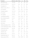
Investigations of blood cells revealed marked leucocytosis in 14 (93%) with a mean of 18.3±5.6×10⁹L–1, neutrophilia and thrombocytopenia in seven cases (47%) with a mean of 164±128×109L–1. Fibrinogen elevation was a typical feature of leptospirosis (mean 5.9±1.7g/L). Laboratory investigations are shown in Table 1.
Laboratory parameters in elderly patients with leptospirosis.
| Parameter | Reference value | Mean | sd | Min | Max |
|---|---|---|---|---|---|
| Hemoglobin (g/L) | 120–188 | 131 | 20 | 100 | 156 |
| Leucocytes (×109L−1) | 4.0–11.0 | 18.3 | 5.6 | 6.1 | 26.4 |
| Neutrophils (%) | 50–80 | 81 | 30 | 67 | 92 |
| Platelets (cells×109L−1) | 150–400 | 164 | 128 | 62 | 437 |
| Urea (mmol/L) | 1.7–8.3 | 38.1 | 24.1 | 11.2 | 98.6 |
| Creatinine (μmol/L) | 44.2–134 | 347.6 | 179.8 | 99 | 610 |
| K+ (mmol/L) | 3.5–5.6 | 4.2 | 1.1 | 2.7 | 6.5 |
| Na+ (mmol/L) | 130–151 | 137 | 5.5 | 129 | 148 |
| Total bilirubin (μmol/L) | 3.4–21 | 274.5 | 210.7 | 35 | 780 |
| Direct bilirubin (μmol/L) | 0.8–8.5 | 219.6 | 155.2 | 27 | 564 |
| ASAT (IU/L) | ≤37 | 126 | 62 | 28 | 625 |
| ALAT (IU/L) | ≤40 | 131 | 127 | 11 | 382 |
| GGT (IU/L) | 15–28 | 238 | 164 | 31 | 508 |
| Alkaline phosphatase (IU/L) | 50–260 | 365 | 334 | 70 | 1431 |
| Lactate dehydrogenase (IU/L) | 100–360 | 1049 | 877 | 287 | 2305 |
| Creatine kinase (IU/L) | 80–190 | 2227 | 3938 | 78 | 8130 |
| Total protein (g/L) | 58–80 | 58.6 | 7.8 | 48 | 72 |
| Albumins (g/L) | 35–55 | 28 | 6.4 | 18.5 | 43.8 |
| Fibrinogen (g/L) | 2.0–4.5 | 5.9 | 1.7 | 3.0 | 8.75 |
| Prothrombin index (%) | 80–110 | 82 | 20 | 47 | 108 |
| Serum amylase (IU/L) | 30–300 | 312.8 | 309.7 | 42 | 1055 |
Fourteen patients (93%) had ARF and one (7%) had prerenal azotemia. Cardiac involvement with clinical and electrocardiographic (ECG) abnormalities was found in eight patients (53%). Four deceased patients had developed myocarditis. Acute respiratory failure due to lung edema was observed in six (40%) patients. Thirteen (87%) had manifested hemorrhagic diathesis such as skin hemorrhage in nine (60%), hematuria in nine (60%), epistaxis in three (20%), hematemesis in two (13%), melena in 1 (7%). Pancreatic involvement was detected in only two (13%) patients.
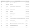
The complex assessment of severity, according to the definitions mentioned above, revealed that 12 cases (80%) had the severe form of leptospirosis and only three cases (20%) had the moderate form. Co-morbidity (hypertonic diseases, chronic pulmonary and heart diseases, chronic alcohol abuse, diabetes, etc.) was reported in 13 cases (87%) (Table 2).
Severity, co-morbidity and outcome of leptospirosis in elderly patients.
| Age (years) | Severity | Co-morbidity | Outcome |
|---|---|---|---|
| 63 | Severe | Hypertonic disease | Survived |
| 60 | Moderate | Blindness | Survived |
| 69 | Severe | Nephrolythiasis, cholelythiasis | Survived |
| 74 | Moderate | None | Survived |
| 68 | Moderate | Pyelonephritis | Survived |
| 71 | Severe | Diabetes | Survived |
| 63 | Severe | Pneumofibrosis | Survived |
| 61 | Severe | Alcohol abuse | Survived |
| 78 | Severe | None | Survived |
| 75 | Severe | Hypertonic disease, ischemic heart disease | Survived |
| 63 | Severe | Stomach ulcer | Died |
| 69 | Severe | Ischemic heart disease | Died |
| 71 | Severe | Myocardial infarction, hypertonic disease | Died |
| 68 | Severe | Ischemic heart disease | Died |
| 75 | Severe | Tuberculosis | Died |
Patient management was initiated immediately after admission and consisted of intravenous administration of fluids and plasma transfusions. Penicillin (200,000U/kg per day) was administered intravenously to twelve patients during 14 days. Intravenous ceftriaxone 2.0–4.0g per day (depending on diuresis and creatinine levels) was administered when penicillin had no effect, due to penicillin allergy or in very severe cases. Steroid was used (methylprednisolone 2mg/kg per day initial dose), famotidine for gastro-protection, furosemide, hepato-protective (l-ornitine, ademethionine). Transfusions of thrombocyte concentrates were carried on in cases with critical thrombocytopenia and severe multi-site bleeding. Human albumin was infused in cases with hypoalbuminemia. Dialysis was performed in three cases, initiated on the second to fourth day because of ineffective conservative treatment of ARF and a totally of two to eight séances were performed.
Despite intensive treatment five cases died (one had been dialyzed). The dynamics of serum bilirubin and creatinine levels in the deceased patients had dramatically increased.
Discussion of the results mentioned above requires analyzing global and local information about leptospirosis. The disease is endemic in both urban and rural areas worldwide and there had been many outbreaks in the recent past.1 Leptospirosis is presumed to be the most widespread zoonosis in the world. The source of infection in humans is usually either by direct or indirect contact with urine of an infected animal. The incidence is significantly higher in warm climate countries than in temperate regions. This is mainly due to longer survival of leptospires in warm and humid environments.2 In Bulgaria, leptospirosis is a reportable disease since 1953, when a database and official registration was initiated. A mean annual incidence rate of 0.9–3.1 per 100,000 was reported during the period 1953–1968 followed by a reduction to 0.1 per 100,000 inhabitants within next ten years. Since 1976, a mean annual incidence of 0.37 per 100,000 was reported.11 A total of 102 leptospirosis cases were treated at the Clinic of Infectious Diseases of University Hospital – Pleven (1976–2012) and only 15 were aged ≥60 years old. As a result only a small sample of elderly patients could be evaluated. Epidemiologic history suggestive of leptospirosis was reported 80% of our cases.
Clinical leptospirosis can vary from a mild non-specific influenza-like infection to a severe disease, where serious complications like ARF, myocarditis, pulmonary hemorrhage and liver failure are reported.2 Case fatality rates have been reported to vary between 3% and 54% as result of the affected organ/system.5 Only 20% of our cases had a moderate course and the remainder had a severe course. Case fatality rate was 33%. Of the deceased patients two had affection of four organ/system, another two had affected five, and one patient had affected six organ/system.
Renal involvement is the most serious complication in leptospirosis and is the commonest cause of death.6 Two mechanisms have been postulated in the production of leptospiral renal failure: (1) direct nephrotoxicity brought about by various endotoxins or endotoxin-like substances; and (2) anoxic effect due to disturbances in renal circulation. The typical lesion is tubulointerstitial nephritis, characterized by interstitial edema and dense focal infiltration of predominantly mononuclear cells. Tubular changes are degenerative in nature and affect principally the proximal tubules. Intravascular volume depletion causing vasomotor nephropathy in this disease has been postulated to occur due to capillary leakage with associated loss of fluid and protein resulting in proteinuria.6 In our study 93% of cases had ARF.
Cardiac involvements are a life-threatening complications of leptospirosis. Rajiv et al. (1996) have shown that 70% of the patients with serologically proven leptospirosis had ECG abnormalities, with atrial fibrillation being the commonest major arrhythmia noted.12 The same study reported that 36% of the patients had conduction system abnormalities and 30% had T-wave changes. Another case-series has found atrio-ventricular block in 44% of patients with leptospirosis.13 A glycoprotein fraction of leptospiral cell wall has been incriminated in the pathogenesis of these rhythm disturbances. Other reported cardiac abnormalities include myocarditis and endocarditis.14 Cardiac involvement with clinical and ECG abnormalities were found in 53% of our cases. Four deceased elderly patients had myocarditis.
We consider that co-morbidity had significant contribution to the severe course observed in elderly patients. All patients who ended up dying had serious co-morbidities that influenced either directly or indirectly the lethal outcome.
Severe leptospirosis may carry a high mortality if treatment is not instituted promptly. Poor prognostic markers in leptospirosis reported in various studies include hypotension, oliguria and hyperkalemia.7 Other studies have reported dyspnea, white blood cell count greater than 12,900mm−3,8 repolarization abnormalities on ECG, alveolar infiltrates,9 hemoptysis, metabolic acidosis, and thrombocytopenia.15 In hospitals that have adequate facilities, patients with these risk factors should preferably be treated in an Intensive Care Unit.4 The beginning of early pertinent antimicrobial therapy within 4–5 days after the onset of illness, proper supportive therapy and use of dialysis to treat renal failure has reduced the leptospirosis-related mortality.4,15 Therefore early predictors of complications are helpful in detecting and managing patients with leptospirosis and also to minimize mortality. In our study 53% of cases were not admitted early to the Clinic of Infectious Diseases due to incorrect primary diagnosis. This is evidence for difficulty to diagnose especially in elderly patients when co-morbidity may overlap the clinical manifestations.
In conclusion, leptospirosis in elderly patients is associated with severe course and higher risk for death and requires prompt intensive treatment.
Conflict of interestThe authors declare no conflict of interest.
