
In the last two decades, the scientific community has recognized the Microsporidia importance and associated it with serious infections in immunocompromised patients.1 Since its discovery in 1857, there was controversies in its phylogenetic classification, being recently classified as fungi.1,2 There have been described 150 genera and over 1300 species. Eight genera are described in human hosts: Enterocytozoon, Encephalitozzon, Pleistophora, Trachipleistophora, Vittaforma, Brachiola, Nosema, and Microsporidium. Fourteen species are associated with human diseases, including Enterocytozoon bieneusi and Encephalitozoon intestinalis. Microsporidia causes systemic and non-systemic diseases, with wide spectrum of clinical manifestation, principally chronic diarrhea.3
As little is known about microsporidia and its importance as infectious agent in immunocompromised patients, we conducted a retrospective study in 347 stool samples analyzed for the presence of microsporidiosis to describe the associated underlying diseases. The data were registered in the Parasitology Laboratory of UNICAMP (2011–2012). All patients for whom there was a medical request to exam the stools for the presence of microsporidia were included. Patients’ underlying disease was recorded.
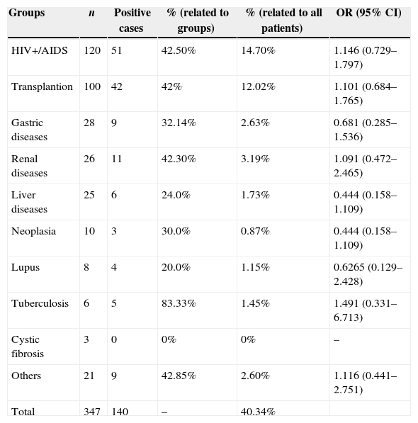
Out of the 347 [210 (60.52%) males] fecal samples analyzed, 140 (40.34%) were positive for microsporidia. The majority of the positive samples came from male patients [94 (67.14%)]. Table 1 shows the underlying diseases of patients found with microsporidia in their feces. The positivity rates were 75% (75/100), 22% (22/100), and 3% (3/100) among those who underwent bone marrow, kidney and liver transplantation, respectively.
Microsporidia identification related to clinical status.
| Groups | n | Positive cases | % (related to groups) | % (related to all patients) | OR (95% CI) |
|---|---|---|---|---|---|
| HIV+/AIDS | 120 | 51 | 42.50% | 14.70% | 1.146 (0.729–1.797) |
| Transplantion | 100 | 42 | 42% | 12.02% | 1.101 (0.684–1.765) |
| Gastric diseases | 28 | 9 | 32.14% | 2.63% | 0.681 (0.285–1.536) |
| Renal diseases | 26 | 11 | 42.30% | 3.19% | 1.091 (0.472–2.465) |
| Liver diseases | 25 | 6 | 24.0% | 1.73% | 0.444 (0.158–1.109) |
| Neoplasia | 10 | 3 | 30.0% | 0.87% | 0.444 (0.158–1.109) |
| Lupus | 8 | 4 | 20.0% | 1.15% | 0.6265 (0.129–2.428) |
| Tuberculosis | 6 | 5 | 83.33% | 1.45% | 1.491 (0.331–6.713) |
| Cystic fibrosis | 3 | 0 | 0% | 0% | – |
| Others | 21 | 9 | 42.85% | 2.60% | 1.116 (0.441–2.751) |
| Total | 347 | 140 | – | 40.34% |
n, number of patients; %, percentage; OR, odds ratio; CI, confidence interval.
The underlying diseases of the positive cases were diarrhea (eight), Crohn's disease (one), leukemia (one), lymphoma (two), chronic renal failure (two), nephritic syndrome (two), end-stage renal disease (five), cirrhosis (six), pulmonary disease (two), immunodeficiency (1), and none (5).
Microsporidiosis is a cosmopolitan disease whose prevalence has wide variability depending on the geographic region, population studied and diagnostic methods.4 Immunocompromised patients are more affected by serious microsporidiosis, especially HIV-patients. In the early ‘80s, before the AIDS epidemic, microsporidiosis was rarely described in humans. Since 1990, the prevalence of HIV-associated microsporidia infection has been reported to range from 1.5 to 50%.3 Transplanted patients are another risk group for microsporidiosis.5
The wide spectrum of clinical manifestations depends on the species, site of infection and host immunity.4 Gastrointestinal microsporidiosis is predominantly observed in HIV-patients, especially with advanced immunosuppression.1
Asymptomatic microsporidiosis has been described in immunocompetent, as well as in immunocompromised patients. The search for microsporidia in fecal samples should be performed in immunocompromised patients, even without symptoms, not only due to the risk of overt symptoms, but also because of the significant transmission risk.1
In conclusion, high positivity for microsporidiosis was observed among HIV-infected patients and transplant recipients, highlighting the importance of early diagnosis and implementation of measures for disease prevention.
Conflicts of interestThe authors declare no conflicts of interest.
