
Results of Chagas’ disease diagnosis show disagreement. The aim of this study was to compare commercial tests for Chagas’ disease serodiagnosis in southern Brazil. A total of 161 samples were evaluated. Three enzyme-linked immunosorbent assays, one indirect hemagglutination and one indirect immunofluorescence were assessed. Trypomastigote excreted-secreted antigen-blot was a confirmatory method. From 161 samples, 65.84% were positive in all tests, while 34.16% presents mismatch result in at least one of the tests. All techniques tested presented false-positive and/or false-negative results as follows: Enzyme-linked immunosorbent assay 1 had more false-positive results (lower specificity), indirect immunofluorescence had the highest rate of false-negative results (lower sensitivity), enzyme-linked immunosorbent assays had fewer false-negative results (higher sensitivity), while indirect hemagglutination showed no false-positive result (higher specificity). Knowing the characteristics of techniques make it possible to combine them and obtain more reliable diagnosis. Therefore, it seems useful to combine techniques for diagnosing this infection.
Chagas’ disease, caused by the protozoan Trypanosoma cruzi, constitutes a serious public health problem in Latin America. Once controlling measures against the triatomine insects are implemented, secondary mechanisms of transmission become relevant, such as blood transfusion, organ transplantation, ingestion of contaminated food and transplacental route, especially in countries where vectorial transmission does not occur.1 In many countries outside Latin America, Chagas’ disease diagnosed among immigrants from endemic areas is a cause for concern.2–5
The diagnosis of Chagas’ disease is difficult, both in routine laboratories dealing with suspected infections and in screening laboratories (blood banks, umbilical cord blood banks, organs donors, etc.). Although there are several tests available to indirect diagnosis (especially enzyme-linked immunosorbent assay (ELISA), indirect hemagglutination (IHA) and indirect immunofluorescense (IIF)), there is not a gold-standard technique, and the results obtained by different techniques show disagreement, with varying levels of sensitivity and specificity.6–8 These results may be related to the parasitic form used to obtain the antigens; there are antigenic differences between epimastigote and amastigote forms, accepting that their immunodominant fractions are not the same.9 Trypomastigotes-based ELISA showed higher specificity10,11 and, in some cases, higher sensitivity too.9 The source of the strain used to obtain the antigen can produce differences in technique performance, with better results with antigens from local strains of T. cruzi.12–14
Nonspecific reactions, causing inconclusive or false positives (FPs) can occur in the diagnosis of Chagas’ disease, most frequently by antibodies produced by patients with leishmaniasis.9,12,15–18 Some confirmatory tests have been described for conventional serology in doubtful situations, such as trypomastigote excreted-secreted antigen-blot (TESA-blot),19 INNO-LIA Chagas (line immunoassay),20 RIPA (radioimmune precipitation assay)21 and Abott Chagas immunoassay.22 TESA-blot shows high sensitivity and specificity in different groups of patients compared to those of conventional serological methods.23,24 It has been used as the gold-standard in the serology of Chagas’ disease.25 TESA-blot is considered positive when sera react with antigens of 130–200kDa and/or antigens of 150–160kDa, and some sera also react with bands of 80–120kDa (SAPA – shed acute-phase antigen). Several chronic patients also react with SAPA bands plus a band of approximately 95kDa.19
This study was conducted to compare different tests utilized for serodiagnosis of Chagas’ disease in patients of Rio Grande do Sul State, Southern Brazil. In order to obtain reliable results for Chagas’ disease it is imperative to understand the performance of the available techniques.
Materials and methodsSample collectionThe tests were evaluated in 161 serum samples from Laboratory of Serology of Blood Center of Pelotas City, Brazil, that were positive in at least one of the techniques utilized in this study. The Blood Center of Pelotas receives blood donors living in both urban and rural areas of south of Rio Grande do Sul State, Southern Brazil. The samples from adults (18–65 years old) were collected and serum samples were frozen at −20°C until the end of analysis.
Ethics statementAt blood collection, all subjects gave written consent to participate in this study. Privacy and anonymity of patients were strictly ensured. The study was approved by the Research Committee of Postgraduate Program of Universidade Federal de Pelotas, Brazil, and by the Blood Center of Pelotas City, Brazil.
Serological methodsELISA 1 – Chagatest® (Wiener Lab, Argentina). ELISA 2 – CHAGATEK® (Biolab-Mérieux, Rio de Janeiro, Brazil). ELISA 3 – EIAgen T. cruzi IgG+IgM (Adaltis, Bologna, Italy). Indirect hemagglutination (IHA) – Chagatest IHA® – Screening AV (Wiener Lab, Argentina). In case of reactive samples, quantitative tests were carried out using the same methodology, performing serial dilutions of serum. Indirect immunofluorescence (IIF) – IMUNOCRUZI® (Biolab-Mérieux, Rio de Janeiro, Brazil) and conjugated sheep anti-IgG human marked by fluorescein isothiocyanate, FLUOLINE® (Biolab-Mérieux, Rio de Janeiro, Brazil).
The tests were evaluated as described in the specific methodology. Positive and negative controls were always included to validate the results.
Confirmatory test – TESA-blotImmunoblotting with antigen TESA (excretory–secretory antigen of T. cruzi) was used as a confirmatory method19,23,24 in samples with discordant results between the techniques and also in reactive samples in all tests, to assess the pattern of bands recognized by sera positive for T. cruzi in patients of southern of Brazil.
Statistical analysisThe Cohen's Kappa index was used to measure the magnitude of agreement beyond chance between serological tests and the confirmatory method TESA-blot. Kappa values >0.81 represent ‘excellent’ agreement; those between 0.61 and 0.8 show ‘good’ agreement; those between 0.41 and 0.6 show ‘moderate’ agreement; those between 0.21 and 0.4 show ‘weak’ agreement and values <0.21 represent ‘negligible’ agreement.
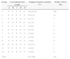
ResultsTable 1 presents the results of 161 serum samples showing reactivity to anti-T. cruzi by the tested techniques. The samples were divided into 12 groups, according to its reactivity in the techniques panel and were compared with TESA-blot results.
Reactivity of 161 samples for Trypanosoma cruzi tested by three ELISAs, an IHA and an IIF compared with TESA-blot result, composing 12 groups, according samples comportment in techniques panel.
| Group | Conventional tests results | Samples frequency number (%) | Positive TESA-blot | ||||
|---|---|---|---|---|---|---|---|
| A | B | C | D | E | |||
| 1 | P | P | P | P | P | 106 (65.84) | 106 |
| 2 | P | P | P | P | N | 10 (6.21) | 10 |
| 3 | P | P | P | N | N | 6 (3.73) | 3 |
| 4 | P | N | P | N | P | 2 (1.24) | 1 |
| 5 | P | P | P | N | P | 1 (0.62) | 0 |
| 6 | P | N | P | N | N | 9 (5.59) | 0 |
| 7 | P | P | N | N | N | 1 (0.62) | 0 |
| 8 | N | P | P | N | N | 4 (2.48) | 0 |
| 9 | N | N | P | N | N | 1 (0.62) | 0 |
| 10 | P | N | N | N | N | 19 (11.8) | 1 |
| 11 | N | P | P | P | P | 1 (0.62) | 1 |
| 12 | N | P | N | N | N | 1 (0.62) | 0 |
| Total | 161 (100) | 122 | |||||
Order of conventional tests: A – ELISA 1, B – ELISA 2, C – ELISA 3, D – IHA and E – IIF. N represents negative results and P represents positive results.
Considering the results of confirmatory testing, 122/161 (75.78%) samples were positive while 39/161 (24.12%) were negative for anti-T. cruzi. The 150–160 and 95kDa bands were showed, in higher or lower intensity, in positive samples by TESA-blot. In some samples, the range of 120–210kDa bands related to SAPA was evident. According to Table 1, from 161 sera analyzed, 106 (65.84%) were positive in all commercial tests (Table 1, group 1). When a sample was positive in all commercial tests, TESA-blot confirmed the result. In the remaining 55 samples (43.16%) mismatch result was observed with at least one of the tests.
All samples showed reactivity to at least one of three types of ELISA tested. ELISAs 1, 2 and 3 had an index of reactivity (OD/cut off) ranging from 0.90 to 8.30, 0.96 to 16.28 and 1.02 to 13.35, respectively. A comparison among the three ELISA tests showed a concordance in 123 samples (76.4%), or even, considering the same methodology, a variation was observed between different types of kits. Reactivity only with one ELISA occurred in 21 samples, which was more common with ELISA 1 (Table 1, groups 9, 10 and 12). Of these, only one sample developed bands with TESA-blot. Positive samples in only two ELISA tests (Table 1, groups 6, 7 and 8) were negative with TESA-blot. Reagent samples with ELISAs and negative with IHA and IIF (6/161) were 50% negative and 50% weakly positive with TESA-blot (Table 1, group 3). Non-reagent sample only with ELISA 1 (Table 1, group 11) was positive with TESA-blot, thus false-negative (FN) results.
With IHA, 117 samples (72.67%) were reactive and 44 (27.33%) were negative. This technique disagreed in isolation with other techniques (Table 1, group 5) in only one sample (0.62%), whose TESA-blot result confirmed IHA result.
In the case of IIF, 110 (68.32%) samples were positive and 51 (31.68%) were negative. The IIF disagreed with other techniques (Table 1, group 2) in 10 samples (6.21%), where IIF showed negative results, whereas the other four techniques and the TESA-blot showed positive results.
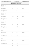
Results with ELISA, IHA and IIF were compared with TESA-blot, highlighting FP and FN results of each technique (Table 2). ELISA 1 had more FP results (33 samples); IIF presented had the highest number of FN results (14 samples); the ELISAs, in general, showed fewer FN results (1 sample with ELISA 1 and ELISA 3, and 2 samples with ELISA 2), whereas there was no FP result with IHA.
Positive and negative cases for each technique compared with TESA-blot (confirmatory method) results and Kappa index.
| Conventional tests | TESA-blot | Kappa index | |
|---|---|---|---|
| Positives | Negatives | ||
| ELISA 1 | |||
| Positives | 121 | 33 | 0.21 |
| Negatives | 1 | 6 | |
| ELISA 2 | |||
| Positives | 120 | 10 | 0.77 |
| Negatives | 2 | 29 | |
| ELISA 3 | |||
| Positives | 121 | 19 | 0.61 |
| Negatives | 1 | 20 | |
| IHA | |||
| Positives | 117 | 0 | 0.92 |
| Negatives | 5 | 39 | |
| IIF | |||
| Positives | 108 | 2 | 0.76 |
| Negatives | 14 | 37 | |
| 122 | 39 | ||
According to Kappa index, the agreement with the confirmatory method was excellent for IHA technique, good for ELISA 2, ELISA 3 and IIF, and weak for ELISA 1.
DiscussionAs observed in this study, the discrepancy between results of serological tests is frequent.7,12,21,24,26–30 A comparison of only ELISA tests in this study showed higher agreement than in other comparative studies.7,8 The variety of T. cruzi antigens used in the preparation of the extract utilized in different serological tests may result in discrepancies in the results when analyzing only one sample. The heterogeneity and genetic variability among strains of T. cruzi are well known, with a predominance of one lineage or population in a location, identified as regional strains, which modifies the biological conditions, virulence, clinical profiles of the host and, consequently, antibodies with variable performance in diagnostic methods.31–36 The use of regional strains provides the best performance compared to the use of strains of various origins.12 Likewise, the parasitic form may interfere with the proven results of serological tests, so that antigens of trypomastigotes, an exclusive form of Trypanosoma, have a better specificity and, in some cases, sensitivity.9,11 In addition to issues related to the kits, other interferences can be responsible for unspecific reactions, such as inadequate samples and biological issues, as cross reactions or individuals on treatment. The most common cross-reactions with Chagas’ disease occur with serum from patients with leishmaniasis,9,18 which was not a problem in this study, since this disease has not been reported in the region of origin of the samples. The subjectivity of the reading techniques of IIF and IHA may also cause variation in samples with weakly reactive results.
All serological techniques tested presented failures for sensitivity and/or specificity, considering the occurrence of FP and/or FN results in all of them (Table 1). The IIF, with highest number of FN, showed more isolated non-reagent results (10/161 samples). In contrast, other studies showed 100% sensitivity with IIF.7 On the other hand, the ELISA techniques in general proved to be more sensitive. The IHA was the technique with the greatest specificity and was the only one with no FP, while the ELISA 1 presented the lowest specificity. Also there was greater specificity with IHA compared to ELISA and, on the other hand, greater sensitivity with ELISA.6
Considering the results of the confirmatory test, when the sample was positive in only one or two techniques, the trend was for a FP result, and the ELISA's reactivity index was low. The same was observed in the confirmatory test INNO-LIA, where the majority of negative results were detected in sera that had reacted in only one of the screening techniques.20
In an attempt to improve the quality of serodiagnosis of Chagas’ disease, it seems useful to combine more than one technique for the diagnosis of this infection. The commercially available diagnostic kits are produced from certain strains of T. cruzi and marketed to different regions. Therefore, it would be worthwhile that at least the antigenic extract used was composed of strains from different regions, with more sensitive and specific parasitic form.9,11 Additionally, a confirmatory test of a higher sensitivity and specificity should be added.23,24
ConclusionThe techniques tested show disagreement in results for anti-T. cruzi detection, confirming the knowledge existing in the literature. All of them presented FP and/or FN results. It is important to know the characteristics of different techniques in order to associate them and obtain a more reliable and appropriate diagnosis to the stage of disease and patient situation.
Conflict of interestThe authors declare to have no conflict of interest.
Our thanks to Dr. Eufrosina Umezawa, from Tropical Medicine Institute of Sao Paulo, Brazil, for providing the TESA-blot used in this study and to PhD Eduardo Xavier, from Universidade Federal de Pelotas, RS, Brazil, for the English review.
