To determine the epidemiological and molecular characteristics of 12 Staphylococcus aureus isolates presenting heteroresistance to vancomycin in laboratories of two cities in Santa Catarina, southern Brazil. Epidemiological data, including the city of isolation, health institution, and date of isolation were considered, as well as the associated clinical specimen. For molecular characterization, we analyzed the staphylococcal cassette chromosome types, the erm gene presence, and the genomic diversity of isolates using pulsed-field gel electrophoresis. The 12 isolates of S. aureus were previously confirmed as heteroresistance to vancomycin using the population analysis profile–area under curve. Regarding genetic variability, two clones were detected: the main one (clone A) composed of four isolates and the clones B, with two isolates. For clone A, two isolates presented identical band patterns and were related to the same hospital, with an interval of 57 days between their isolation. The other isolates of this clone showed no epidemiological link between them because they were isolated in different hospitals and had no temporal relationship. The other clone showed no detectable epidemiological relationship. The heteroresistance to vancomycin recovered in Santa Catarina State from 2009 to 2012 had, in general, heterogeneous genomic patterns based on pulsed-field gel electrophoresis results, which is in accordance with the fact that these isolates had little or no epidemiological relationship among them. Due to the characteristic phenotypic instability and often prolonged vancomycin therapy for selection, clonal spread is not as common as for other resistance mechanisms disseminated through horizontal gene transfer.
Staphylococcus aureus can acquire, during prolonged therapy with glycopeptides, a very peculiar resistant phenotype. Through selective pressure and apparently without transfer of genetic material, it may undergo mutations in genes responsible for cell wall production, making it thicker and less susceptible to the action of antimicrobials.1
The heteroresistance of S. aureus to vancomycin (hVISA) causes changes in the macro-morphological features of the colonies, that present with heterogeneous appearance and pigmentation, giving the impression of contamination and may confuse the microbiologist.2 Its mechanism of resistance is associated with activation of cell wall synthesis, which increases the production of waste mucopeptide and reduces the amount of antibiotic that reaches the site of action, thus causing cell wall thickening and subsequent drug imprisonment.3 Speculation that hVISA could be regarded as precursors of VISA strains once, after prolonged exposure to antimicrobial could select a homogenous population of cells expressing the phenotype VISA. This phenotype is unstable.4
The detection of infections caused by hVISA represents a challenge for microbiologists, since these strains are considered susceptible to vancomycin in vitro (minimum inhibitory concentration (MIC) ≤2μg/mL), and therefore categorized as susceptible by the usual laboratory methods.1 However, as it contains subpopulations 1 in each 106 bacterial cells, that can grow in the presence of 4μg/mL of vancomycin, it may lead to treatment failure5,6 with vancomycin,.
Reference methods used to evaluate susceptibility as broth microdilution, Etest and automated methods fail to detect hVISA. Because the phenotype is a heterogeneous phenomenon, reliable molecular markers to confirm this phenotype have not yet been found. Partly due to the small inoculum, relatively poor growth on Mueller-Hinton agar, or incubation for only 24h. The inoculum size is critical to the detection of subpopulation of resistant cells; furthermore hVISA strains are notoriously slow growing, with cell walls thicker and pleomorphic unique characteristics, with colonies of varying sizes and nutritionally exacting.7
The population analysis profile–area under the curve (PAP–AUC) has been the most reliable and reproducible approach, being considered the gold-standard test for hVISA.8 It was specifically designed for discriminating hVISA and VISA. It is a method of analysis of modified sub-populations using serial concentrations of vancomycin, in order to quantify the viable bacterial populations in such concentrations. It is a very laborious, expensive, and inappropriate for routine use in clinical laboratories.9
In a meta-analysis published in 2011, the rates of treatment failure (designated as persistent infection or bacteremia) related to hVISA isolates were two times more common than in infections caused by S. aureus susceptible to vancomycin (OR: 2.37, 95% CI: 1.53–3.67).6 Therefore, an accurate and practical laboratory method for the detection of hVISA isolated in clinical practice is of increasing importance.10
Despite the controversy between studies regarding the association of hVISA and mortality, knowledge of the epidemiological profile is very important in assisting the clinician when choosing the appropriate antibacterial therapy. The objective of this study was to evaluate the phenotypic and molecular epidemiology characteristics of hVISA isolates in the state of Santa Catarina, Brazil.
MethodsBacterial samplesWe used 12 clinical isolates of hVISA obtained from various anatomical sites of patients in hospitals in Florianópolis and hospital in Blumenau, all located in the state of Santa Catarina in southern Brazil. Samples were collected from November 2009 to October 2012.
Antimicrobial susceptibility testingAntimicrobial susceptibility testing was performed using the disk diffusion method, according to the recommendations and interpretive criteria of the Clinical and Laboratory Standards Institute.11 We also performed the D test for the detection of inducible resistance to clindamycin. Disks of erythromycin and clindamycin were placed 26mm apart, and a flattening of the inhibition zone indicated a positive test, which was reported as clindamycin resistance. Vancomycin MICs were determined by macrodilution method.11
PAP–AUCAfter incubation on solid medium, the bacteria were diluted in sterile saline at dilutions ranging from 10−1 to 10−8 and subsequently spotted as 10-μL spots on BHI agar plates containing 0, 0.5, 1, 2, 3, 4, 5, 6 and 8μg/mL of vancomycin. The plates were incubated for 48h, and the colonies were counted to determine the log10 CFU/mL; these data were then plotted on a graph as a function of the vancomycin concentration. The AUC was calculated using the strain Mu3 (ATCC 700698) as a control. To confirm the designation as hVISA, the ratio of the AUC of the isolate to that of the Mu3 strain must be greater or equal to 0.9 and non-hVISA isolates had a PAP–AUC<0.9.7,8
Multiplex PCR for the detection of the staphylococcal cassette chromosome (SCCmec)The SCCmec type was determined using the multiplex PCR method according to the protocol developed by Zhang et al. The amplicons that were formed had the following sizes: I (613bp), II (398bp), III (280bp), IVa (776bp), IVb (493bp), IVc (200bp), IVd (881bp), and V (325bp).12,13
PCR for erm gene detectionFor isolates with positive results in the phenotypic test for inducible resistance to clindamycin, erm gene PCR amplification was performed according to the multiplex PCR protocol developed by Khan et al.14 The PCR products (610bp for ermA and 520bp for ermC) were analyzed by electrophoresis through a 1.5% agarose gel.14,15
Pulsed-field gel electrophoresis (PFGE)PFGE was performed according to McDougal et al.16 and Pinto et al.17 The fragments were subjected to PFGE using 1% agarose gels (Pulsed Field Certified Agarose; Bio-Rad) in 0.5× Tris-borate-EDTA buffer with a CHEF-DR III system (Bio-Rad). The gels were stained with 0.5μg/mL ethidium bromide, visualized under UV light, and photographed using a GelDoc™ XR System (Bio Rad). The PFGE patterns were analyzed using Bionumerics version 6.1 (Applied Maths, Sint-Martens-Latem, Belgium). The PFGE patterns were clustered by UPGMA. A dendrogram was generated from a similarity matrix calculated using the Dice similarity coefficient with an optimization of 0.5% and a tolerance of 1%. PFGE clusters were defined as isolates with a similarity of 80% or higher on the dendrogram.18
ResultsThe twelve hVISA isolates were recovered from tracheal aspirates (n=5), osteomyelitis (n=4), blood (n=1), skin lesion (n=1), and surgical wound (n=1). Resistance of the 12 hVISA isolates to antimicrobial agents was assessed by disk diffusion, with the following results: clindamycin (92.7%), erythromycin (100%), trimethoprim/sulfamethoxazole (16.7%), ciprofloxacin (92.7%), tetracycline (16.7%), chloramphenicol (8.3%), and gentamicin (33.3%). All isolates were considered susceptible to linezolid and teicoplanin. The vancomycin MICs were 1.0μg/mL (33.3%) and 2.0μg/mL (66.7%) (Fig. 1).
Growth curves of the twelve isolates characterized as hVISA compared to the standard strain Mu3. All isolates showed an AUC/Mu3 AUC ratio greater than 0.9. For clarity, bacterial growth was expressed in log10 CFU/mL. Nine concentrations of vancomycin (0, 0.5, 1.0, 2.0, 3.0, 4.0, 5.0, 6.0 and 8μg/mL) were used. Graph A depicts isolates S4, S11 and S13. Graph B shows isolates L10, L43 and L36. Graph C shows isolates L54, L69 and L74. Graph D shows isolates L80, L84 and L92. The reference strain Mu3 is shown in all graphs.
Only two isolates (10 and 54) exhibited inducible clindamycin resistance, as determined by a positive D test. These isolates were subjected to PCR for erm gene detection, and both contained the ermA gene. Isolate 84, which was resistant to erythromycin and sensitive to clindamycin, yielded a negative D test, and the erm gene was not amplified. All other isolates showed constitutive clindamycin resistance.
Isolate SI4 contained two SCCmec types (I and II). Isolate 84 contained SCCmec type I. Isolates S11, 36, 54, 69, 80 and 92 contained SCCmec type II. SCCmec type III was observed in isolates SI13, 10 and 43. Although isolate 80 was considered to be a community isolate, it did not contain SCCmec type IV, which is frequently associated with community-acquired methicillin-resistant S. aureus (CA-MRSA). Only the isolate 74 showed SCCmec type IV (IVc). The isolation of the microorganism in the first 48h of admission was used as the criterion for classification as CA-MRSA.
PFGE was used to assess the degree of genetic similarity, and the results are presented in Fig. 2. Two clones were observed: a primary clone (A), comprising four isolates (SI4, L54, L69, and L92); and clone B, comprising isolates L36 and L80. Isolates SI11, SI13, L10, L43, L74, and L84 did not display genetic similarity with any of the other isolates. The criteria established by Tenover were used for classification, with a similarity index of 80%.
Dendrogram showing the similarities between the 12 hVISA isolates. Two clones were observed: one comprising four isolates (A), two of which displayed 100% similarity (L54 and L69); and clone B, which comprised two isolates that were genetically indistinguishable (L36 and L80). The SI11, SI13, L10, L43, L74 and L84 isolates did not exhibit any similarity with the other isolates.
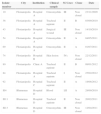
In addition to the molecular characteristics described above, Table 1 presents the city of isolation, attended institution, biological sample, and isolation date. The samples were isolated in two different cities that are 120km apart. In the city of Florianópolis, the state capital of Santa Catarina, three hospitals and two clinics were included. Approximately 33 months separated the first and last isolates. Isolates L54 and L69 were isolated in the same hospital, had 100% genetic similarity, and were isolated from patients with osteomyelitis approximately 57 days apart, which might indicate a common route of infection.
Epidemiological and clinical characteristics of the 12 hVISA isolates.
| Isolate number | City | Institution | Clinical sample | SCCmec | Clone | Date |
|---|---|---|---|---|---|---|
| 10 | Florianópolis | Hospital A | Osteomyelitis | III | Non clonal | 23/11/2009 |
| 36 | Florianópolis | Hospital A | Tracheal aspirate | II | B | 03/09/2010 |
| 43 | Florianópolis | Hospital A | Surgical wound | III | Non clonal | 14/10/2010 |
| 54 | Florianópolis | Hospital A | Osteomyelitis | II | A | 04/05/2011 |
| 69 | Florianópolis | Hospital A | Osteomyelitis | II | A | 01/07/2011 |
| 74 | Florianópolis | Hospital B | Skin lesion | IVc | Non clonal | 22/12/2011 |
| 80 | Florianópolis | Clinic A | Tracheal aspirate | II | B | 09/01/2012 |
| 84 | Florianópolis | Hospital A | Tracheal aspirate | I | Non clonal | 25/02/2012 |
| 92 | Florianópolis | Hospital C | Tracheal aspirate | II | A | 18/08/2012 |
| SI4 | Blumenau | Hospital D | Blood | I,II | A | 28/09/2010 |
| SI11 | Blumenau | Hospital D | Tracheal aspirate | II | Non clonal | 20/02/2011 |
| SI13 | Blumenau | Hospital D | Osteomyelitis | III | Non clonal | 12/04/2011 |
As far as we know, this is the first description of hVISA in Brazil. Two Brazilian studies reported decreased susceptibility to vancomycin, but have not confirmed hVISA phenotype. In 2001, Oliveira and colleagues reported five clinical isolates of MRSA with MIC of 8μg/mL, which were named VRSA, based on available criteria (NCCLS-1997). However, the isolates did not harbor Van genes and, as previously demonstrated by other authors, presented a cell wall thickening.19 In 2006, Lutz and Barth described 18 clinical isolates as possible hVISA, since they were positive for screening test (BHI with 4μg/mL of vancomycin). However, the PAP–AUC confirmatory test for hVISA was not performed, especially because, on that time, the current accepted criteria for confirmation of this phenotype were not known.20,21 Based on these data, we believe our study provides the first report of hVISA isolation in Brazil.
The most frequently anatomical sites associated with hVISA infections are those with a higher bacterial inoculum (abscesses, pneumonia) and those associated with chronic infections (endocarditis, osteomyelitis) for which the use of vancomycin for prolonged periods is quite common and the low penetration of the antibiotic in these sites favors the development of resistance.7 Several studies3,22–25 have established that blood, lower respiratory tract, skin wounds, abscesses and osteomyelitis are the most common sites of hVISA isolation. These findings are similar to those of the present study.
The isolates exhibited a profile of heterogeneous susceptibility, characterized by low resistance to chloramphenicol and trimethoprim/sulfamethoxazole, drugs that are used infrequently to treat MRSA infections. Other studies22,24 have reported low rates of resistance to sulfonamides in hVISA isolates, ranging from 9 to 9.9%. By contrast, studies of samples from Korea26 and China23 revealed much higher values, 58 and 54%, respectively. A chloramphenicol resistance rate of 16.7% was observed in our study, while Hu and colleagues observed a rate of 47.6% in 2013.23 Resistance rates to macrolides, lincosamides, aminoglycosides, fluoroquinolones, and tetracyclines are high, with values greater than 64%, precluding its empirical use.22–24,26 Knowledge of the local susceptibility profile is essential for adequate empirical therapy.
Clindamycin is a therapeutic option for the treatment of serious infections caused by S. aureus, including MRSA, particularly for isolates with SCCmec type IV, which usually are resistant only to beta-lactams.27 However, one common mechanism of resistance is the macrolide-lincosamide-streptogramin B (MLSB) phenotype, which expresses inducible clindamycin resistance, which cannot be detected by traditional phenotypic tests and requires the D test.28 In our study, two isolates exhibited inducible clindamycin resistance, and both possessed the ermA gene. Both were reported to be resistant to clindamycin, and thus inappropriate treatment was avoided.
There was a predominance of SCCmec type II, which is associated with hVISA; in MRSA, this type is associated with higher mortality rates.23 Other studies have also demonstrated a predominance of SCCmec type II among hVISA isolates.22,23,29,30 Some studies have demonstrated a predominance of other types of SCCmec between hVISA isolates such as type I31 and type III.32–34 It is interesting to note that among the 12 isolates, only one had SCCmec type IV (IVc). Although some studies did not find type IV SCCmec among their MRSA isolates,31,34 others demonstrated rates as high as 16.8%22 to 26.3%.32
From an epidemiological perspective, it is difficult to perform further analyses due to the geographical distance and the time interval between the isolation of the first and last isolates. The microorganisms were isolated over a period of three years in different cities and even in several different institutions in the city of Florianópolis. Except for isolates L54 and L69, which are the same clone and were isolated in the same institution, the hVISA isolates did not have any characteristics that would enable an epidemiological link to be made between them.
The hVISA recovered in Santa Catarina State from 2009 to 2012 had, in general, heterogeneous genomic patterns according to the PFGE results, which is in accordance with the fact that these isolates had little or no epidemiological relationship among them.
It should be noted that due to its continental dimensions and tourist vocation, Brazil presents a great diversity of resistance mechanisms, and S. aureus with resistance to vancomycin (VRSA) was first described in 2014 in South America.35 This fact demonstrates the real need for the surveillance of bacterial resistance in our country.
Epidemiological information from each health facility, as well as each geographical region, is critical to the implementation of appropriate empirical therapy, especially with regard to hVISA isolates. Such bacteria have phenotypic characteristics that are difficult to be correctly identify by conventional methods (MIC determination and molecular tests) and are associated with worse clinical outcomes. Due to the characteristic instability phenotype and often prolonged vancomycin therapy for selection, clonal spread is not as common as for other resistance mechanisms disseminated through horizontal gene transfer.
It is imperative that clinical microbiology laboratories detect the hVISA phenotype and associate it with the epidemiological characteristics of the patients (e.g., age, date of isolation, anatomical site, prior use of vancomycin) to provide important information for research laboratories. Through appropriate methodologies, associations between the microbiological data and the clinical characteristics of patients may enable the detection of possible associated risk factors and aid in assessing the prognosis of patients.
Conflicts of interestThe authors declare no conflicts of interest.
The authors thanks to Coordenação de Aperfeiçoamento de Pessoal de Nível Superior (CAPES), Conselho Nacional de Pesquisa e Desenvolvimento Tecnológico (CNPq) and Fundação de Amparo à Pesquisa do Rio Grande do Sul (FAPERGS) for financial support. Thanks to Laboratório Santa Luzia (Cassia Zoccoli and Nina Tobouti) and Laboratório Santa Isabel (Marcelo Molinari) for the granting of clinical samples.


