Lower airway infection is a major cause of morbidity and mortality in patients with cystic fibrosis. It is currently unknown if the infection of the upper airway can cause exacerbation of lower respiratory tract infection. This study aimed to determine the microbiological profile of the anterior paranasal sinuses outflow tract (middle meatus) of cystic fibrosis outpatients. The microbiological profile was defined using endoscopically directed middle meatal cultures. Paired middle meatal and sputum specimens were collected from 56 outpatients for aerobic cultures. A semi-quantitative leukocyte count of the middle meatal samples was performed. The median age of patients was nine years (3–20 years). Staphylococcus aureus (37%), Staphylococcus coagulase-negative (25%), Neisseriac (14%), Pseudomonas aeruginosa (11%), and Streptococcus pneumoniae (7%) were the most prevalent microorganisms in the middle meatal cultures. Using the middle meatal leukocyte count, 16 out of 54 patients (29.6%) presented sinus infection. The most frequently identified pathogens in patients with sinus infections were Staphylococcus aureus (10 patients), Pseudomonas aeruginosa (4 patients), and Streptococcus pneumoniae (3 patients). Agreement of paired middle meatal and sputum cultures was significantly higher among patients with infection in middle meatal (69%). The most common middle meatal pathogens were the typical cystic fibrosis spectrum. This suggests the potential for participating in post-nasal lower airway seeding.
Cystic fibrosis (CF) is the most frequent autosomal recessive disorder of European descent populations, with an incidence of 1:2000–1:8000 live newborns.1,2 Mutations of the CF transmembrane conductance regulator (CFTR) gene lead to a defective secretion of chloride and a hyperabsorption of sodium. This results in depletion of airways surface liquid, which creates abnormally thick and viscous mucus. This mucus is responsible for the reduction in the mucociliary clearance and susceptibility to airways infection and inflammation seen in patients with CF.3–5
CF is a multisystemic disease. Patients commonly present with chronic upper and lower airways infection. Lower respiratory tract infection is a major cause of morbidity and mortality in patients with CF.5Pseudomonas aeruginosa is the most prevalent bacteria in death-related pulmonary infection.6–8 It is also a common pathogen in chronic rhinosinusitis (CRS).9 In patients with CF, radiographic evidence of sinusitis is almost universally detected. Due to persistent symptoms, CRS affects the quality of life of CF patients. Additionally, it may be implicated in increased pulmonary exacerbations, and probably acts as a bacterial reservoir for chronic drainage to lower airways. Colonization of both the upper and inferior respiratory tract with identical Staphylococcus aureus and Pseudomonas aeruginosa has been investigated in previous studies and there is potential for cross-infection between these two sites.9–12
Optimal treatment strategies for bacterial infection in CF depend upon reliable detection of pathogens. In contrast to pulmonary infection, antimicrobial therapy for CRS in CF patients is usually determined empirically. This is especially common in pediatric populations, because there is limited application of the gold standard maxillary sinus puncture. Endoscopically directed middle meatal (MM) culture is an alternative in acute and CRS.13,14 It represents not only the maxillary, but also all anterior sinuses outflow.
At this time, few studies have investigated the MM microbiological flora in CF.15 There is no consensus in the field as to whether the bacteria from the upper airways are implicated in lower respiratory tract infections of these patients. This study aims to determine the microbiological profile of the anterior paranasal sinuses outflow tract (MM) of CF outpatients younger than 21 years of age. A better understanding of microbial flora, an important component of CF disease, can lead to improved diagnostic and treatment strategies.
A prospective study evaluating consecutive CF outpatients receiving care at the Pediatric Pulmonary Clinic of Bahia Reference Center for CF, in Octávio Mangabeira Hospital, was conducted from December 2007 to Jun 2008. Paired MM and sputum samples were collected from study participants, who were also submitted to paranasal sinus computed tomographic (CT) scan. All patients or parents completed a standard questionnaire, which provided information on demographics, signs and symptoms of rhinosinusitis, associated pulmonary disease and history of hospital admission.
Inclusion criteria were diagnosis of CF confirmed by sweat test or genotype and age ranging from three to 20 years. Patients were excluded if they had a history of nasoenteral feeding, antibiotic, oral or nasal corticosteroid therapy in the last 30 days. Those patients who did not tolerate nasal endoscopic examination were also excluded. Informed consent was obtained from all patients prior to examination. The study was approved by the Institutional Review Boards of the Oswaldo Cruz Foundation, Brazilian Ministry of Health.
Two MM samples per patient were collected unilaterally in all cases under endoscopic guidance: one for culture and the other for cytological analysis. Routinely, the turbinates, MM, sphenoethmoidal recess and nasopharynx were assessed for the presence of polyps, purulent secretion, and adenoid hypertrophy. The method was preformed, as follows: nasal mucosa were sprayed with 0.25–0.5% oximetazoline and 2% xylocaine solutions using an atomizer. Endoscopy was performed by retracting nasal ala using a 2.7-mm 30° telescope. Sterile cotton-tipped wire swabs (sterile nasopharyngeal calcium alginate tipped applicators; Puritan Products Co., Guilford, ME) were carefully placed into the MM near the natural maxillary ostium, and turned until the fibers got wet. In order to decrease sample contamination with flora from vestibular, septum or turbinate areas, decongestion of nasal mucosa was performed and the passage of swab was guided by endoscopy. The first swab was placed into the Stuart transport medium and sent to the clinical microbiology laboratory. The other was used to perform a smear on two macroscopic slides, which were fixed with 70% alcohol and sent to the pathology laboratory. Sputum samples were collected by the physiotherapist, during routine respiratory physical therapy, or immediately after the conclusion of otolaryngologic procedures, Sputum and MM samples were immediately sent to microbiological analysis.
Chocolate agar, blood agar, MacConkey agar, Sabouraud Dextrose agar, and Burkholderia cepacea agar base (oxoid) were inoculated with the samples and incubated for 18–48h at 36°C. Mycological analysis was performed by direct examination and culture in Sabouraud medium with chloramphenicol and cycloheximide (BBL). The cultures were incubated at room temperature for at least three weeks.
The number of polymorphonuclear leukocytes (PMNs) in the MM samples was determined by a semiquantitative method, using hematoxylin and eosin (H&E) staining procedure. Smears were examined under oil immersion objective at 1000× magnification independent of the cultures results. PMNs were counted in 20 oil immersion fields, and the average number of PMNs per field was calculated. The results were classified as follows: many (≥10PMNs/field), few (6–10PMNs/field), rare (1–5PMNs/field), none (0PMN/field).
In addition to the paired MM and sputum samples microbiological evaluation, all patients were submitted to sinus CT scan. Three constant anatomic-functional structures, in correspondence to MM microbiological evaluation during this study, were chosen for description: maxillary, anterior ethmoid sinuses and ostiomeatal unit. The degree of opacification at these sites was classified according to Lund–Mackay score. Scores for maxillary and ethmoid sinus were 0 – no opacification, 1 – partial opacification and 2 – total opacification. For the ostiomeatal unit was scored as 0 – no opacification and 2 – total opacification. A Toshiba 64-canals multidetector unit was used to perform axial sections and multiplanar reconstructions without intravenous contrast of all patients. A radiologist and the main investigator analyzed the images.
McNemar's test was used to assess the significance of the difference between two paired proportions. The chi-square test was used for the comparison of categorical variables. Statistical significance was determined at the 5% (p<0.05) level. Statistical analysis was performed using SPSS 14.0 (SPSS, Inc., Chicago, IL).
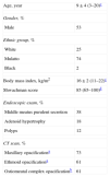
Fifty-six CF patients were evaluated during the study period. The median age was nine years, ranging from three to 20 years (mean±SD, 9±4 years). Fifty three percent of patients were male, 25% white, 74% mullato, and 2% black. The body mass index ranged from 11 to 22 (mean, 16). The median for Shwachman score was 85 (range, 65–100). At endoscopic examination, 38% presented purulent MM secretion, 18% had obstructive adenoid hypertrophy and 12% had nasal polyps. Opacification of maxillary sinus was present in 73% of cases. Ethmoid sinus and ostiomeatal unit were opaque in 61% of patients (Table 1). Severe edema of MM was observed in 18% of patients.
Demographic and clinical findings of 56 cystic fibrosis patients.
| Age, year | 9±4 (3–20)c |
| Gender, % | |
| Male | 53 |
| Ethnic group, % | |
| White | 25 |
| Mulatto | 74 |
| Black | 2 |
| Body mass index, kg/m2 | 16±2 (11–22)c |
| Shwachman score | 85 (65–100)d |
| Endoscopic exam, % | |
| Middle meatus purulent secretion | 38 |
| Adenoid hypertrophy | 18 |
| Polyps | 12 |
| CT scan, % | |
| Maxillary opacificationa | 73 |
| Ethmoid opacificationa | 61 |
| Ostiomeatal complex opacificationb | 61 |
Note. CT, computed tomography.
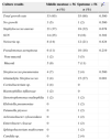
Out of 112 cultures (paired specimens – MM and sputum), 108 (96%) were positive with a total of 147 isolates. The most frequent bacteria were S. aureus, CoNS, Neisseria sp., P. aeruginosa, and S. pneumoniae in both MM and sputum samples. The exception was α-hemolytic Streptococcus, which was more commonly isolated in sputum cultures (p=0.001) (Table 2). Gram-negative bacteria were isolated in 29% of the MM cultures and in 50% of sputum samples. Based on PMN counts in MM samples, 17% of patients presented many, 13% had few, 61% had rare, and 9% did not present any PMNs. Thirty percent of patients (16/54) had more than six PMMs per field. These patients were most frequently colonized by S. aureus (62%), P. aeruginosa (25%) and S. pneumoniae (18%). In patients with rare PMNs, the most frequent bacteria were CoNS (12/38), S. aureus (11/38), Neisseria sp. (6/38) (Table 3). The most frequent bacteria isolated in the MM secretion from patients with severe edema in MM were S. aureus (50%) and S. pneumoniae (30%).
Middle meatal and sputum microbiological profile of 56 cystic fibrosis patients.
| Culture results | Middle meatusn=56 | Sputumn=56 | p* |
|---|---|---|---|
| n (%) | n (%) | ||
| Total growth rate | 53 (95) | 55 (98) | 0.500 |
| No growth | 3 (5) | 1 (2) | 0.500 |
| Staphylococcus aureus | 21 (37) | 18 (32) | 0.678 |
| SCN | 14 (25) | 9 (16) | 0.302 |
| Neisseria sp. | 8 (14) | 12 (21) | 0.424 |
| Pseudomonas aeruginosa | 6 (11) | 10 (18) | 0.219 |
| Non-mucoid | 1 (2) | 3 (5) | |
| Mucoid | 5 (9) | 7 (11) | |
| Streptococcus pneumoniae | 4 (7) | 2 (4) | 0.500 |
| α-hemolytic Streptococcus | 2 (4) | 15 (27) | 0.001 |
| Corinebacterium sp. | 2 (4) | 0 | |
| Haemophillus influenzae | 1 (2) | 0 | |
| Stenotrophomonas maltophilia | 1 (2) | 1 (2) | |
| Klebsiella pneumoniae | 0 | 1 (2) | |
| Tatumella ptyseos | 0 | 1 (2) | |
| Achromobacter xylosoxidans | 0 | 1 (2) | |
| Enterobacter cloacae | 0 | 1 (2) | |
| Sphingobacterium multivorum | 0 | 1 (2) | |
| Candida sp. | 0 | 2 (4) | |
CoNS, coagulase-negative Staphylococci.
Polymorphonuclear leukocyte count from MM material from 56 patients with cystic fibrosis.
| Bacterial species | PMMs account | |
|---|---|---|
| Many or fewn=16 (%) | Rare or nonen=38 (%) | |
| Staphylococcus aureus | 10 (62) | 11 (29) |
| Pseudomonas aeruginosa | 4 (25) | 2 (5) |
| Streptococcus pneumoniae | 3 (18) | 1 (3) |
| CoNS | 2 (12) | 12 (32) |
| Neisseria sp. | 0 | 6 (16) |
| α-hemolytic Streptococcus | 0 | 2 (5) |
| Corynebacterium sp. | 0 | 2 (5) |
| Haemophillus influenzae | 0 | 1 (3) |
| Stenotrophomonas maltophilia | 0 | 1 (3) |
| No growth | 0 | 3 (8) |
PMNs, polymorphonuclear leukocytes; CoNS, coagulase-negative Staphylococci.
Two patients were excluded.
The present study was the first to evaluate the microbiologic flora of the MM in CF outpatients from Brazil using an endoscopically directed MM sample collection. The invasive gold standard maxillary sinus culture puncture is not routinely used in clinical practice. Compared to methods involving direct sinus cavity assessment (endoscopy-guided sinus aspiration, sinus puncture and intraoperative sampling), endoscopic culture may be considered a non-invasive procedure.
S. aureus was the most frequent pathogen, followed by CoNS, Neisseria sp., P. aeruginosa and S. pneumoniae. Gram-negative bacteria accounted for nearly one third of the total samples. Similarly, a high frequency of S. aureus and P. aeruginosa isolated in sputum of CF patients, mainly in patients below 17 years old was described. Methicillin resistant S. aureus was isolated from 6% of these patients, indicating that colonization of respiratory tract by pathogenic microorganisms of these patients occurs at young age.16 The MM microbiological profile found in this study differed from both the maxillary sinus flora of CF pediatric surgical patients and the MM aspirates of a CF general population, in which greater rates of P. aeruginosa and H. influenzae were found.15,17 In fact, surgical patients frequently have more severe chronic sinus disease, with persistent P. aeruginosa infection.18 Likewise, this organism is more frequently isolated in older patients with purulent MM secretion.15 Patients enrolled in the present study were younger than 21 years old, followed in outpatient clinic and had no clinical signs of acute CF exacerbation. Furthermore, such differences in the microbiological profiles are unlikely to be explained by the sampling method, because there is a high concordance rate between maxillary sinus and endoscopically directed MM cultures.13,14
About one third of included patients presented MM purulent secretion and positive culture to S. aureus, P. aeruginosa and S. pneumoniae. In contrast, ostiomeatal complex opacification at CT scans was found in two thirds of the cases. The CT may overestimate the diagnosis of sinus disease and cannot accurately differentiate between mucosal thickening and infection.19
Comparing pathogen profile from MM and sputum cultures, the most frequent isolated bacteria were similar. The exception was seven-fold higher frequency of α-hemolytic Streptococci in sputum culture as compared to MM culture. This may represent sample contamination with oropharyngeal microbiota, especially because the studied patients were composed of young children and it is difficult to collect spontaneous sputum samples. The agreement rate of bacterial profile was higher in the group of patients with MM infection. This may reflect the post-nasal lower airway colonization by pathogenic bacterial species. The genotypic characterization of isolates from both MM and sputum samples could confirm the traffic of pathogenic bacteria from upper to lower airways. There is a high genotypic concordance of P. aeruginosa and S. aureus isolates from nasal and sputum samples, suggesting that the flow of pathogenic bacteria from upper to lower airways may be a frequent event.12 Moreover, it was demonstrated that there is no significant differences in the bacterial community diversity in lung between baseline and exacerbation samples of patients with CF.20
One of the limitations of this study was its cross-sectional design, limiting the understanding of the complex dynamic characteristic of chronic infections in CF patients. In the future, prospective studies should be conducted to define the impact of sinus infection on pulmonary disease in CF patients. In conclusion, the most common MM pathogens in CF young outpatients were the typical CF spectrum and S. pneumoniae. This finding suggests the potential for post-nasal lower airway seeding.
Financial supportSistema Único de Saúde (SUS), Brazilian Ministry of Health.
Conflicts of interestThe authors declare no conflicts of interest.
This manuscript is part of Alessandro Tunes Barros's M.Sc. thesis, Bahia School of Medicine, Postgraduate Course in Medicine and Human Health. We thank all professionals of the Pediatric Pulmonary Clinic of Bahia Reference Center for Cystic Fibrosis, in Octávio Mangabeira Hospital, Brazil for their wonderful collaboration during the study and Tamy Fagundes for her efficient help during microbiological collection and analysis.


