There is scarce information regarding clinical evolution of HBV infection in renal transplant patients.
AimsTo evaluate the prevalence of acute exacerbation in HBV-infected renal transplant patients and its association with the time after transplantation, presence of viral replication, clinical evolution, and use of antiviral prophylaxis.
Materials and methodsHBV infected renal transplant patients who underwent regular follow-up visits at 6-month intervals were included in the study. The criteria adopted to characterize exacerbation were: ALT >5× ULN and/or >3× baseline level. Predictive factors of exacerbation evaluated were age, gender, time on dialysis, type of donor, post-transplant time, ALT, HBeAg, HBV-DNA, HCV-RNA, immunosuppressive therapy, and use of antiviral prophylaxis.
Results140 HBV-infected renal transplant patients were included (71% males; age 46±10 years; post-renal transplant time 8±5 years). During follow-up, 25% (35/140) of the patients presented exacerbation within 3.4±3 years after renal transplant. Viral replication was observed in all patients with exacerbation. Clinical and/or laboratory signs of hepatic insufficiency were present in 17% (6/35) of the patients. Three patients died as a consequence of liver failure. In univariate analysis variables associated with exacerbation were less frequent use of prophylactic/preemptive lamivudine and of mycophenolate mofetil. Lamivudine use was the only variable independently associated with exacerbation, with a protective effect.
ConclusionsAcute exacerbation was a frequent and severe event in HBV-infected renal transplant patients. Prophylactic/preemptive therapy with antiviral drugs should be indicated for all HBsAg-positive renal transplant patients.
According to the World Health Organization, the number of chronic hepatitis B virus (HBV) carriers exceeds 350 million worldwide.1 Among renal transplant patients HBV infection continues to be an important cause of morbidity and mortality, although its incidence declined after the introduction of hepatitis B vaccine in 1982 and as a result of improved overall care during hemodialysis.2 The prevalence of chronic hepatitis B after kidney transplantation ranges from 2 to 21% according to geographic regions.3
Although data about the natural course of HBV infection in renal transplant recipients are scarce, evidence indicates that viral replication is accelerated by immunosuppression and that HBV-related liver disease is more aggressive in renal transplant recipients.4 Some studies have demonstrated that the progression of liver disease could occur in more than 80% of HBsAg-positive renal transplant patients, with a high mortality rate5 and higher incidence of graft loss.6
Reactivation of HBV infection in immunosuppressed patients can be separated into three phases: (1) increase in HBV replication; (2) appearance of hepatic injury (ALT flares) and (3) recovery.7 Biochemical evidence of reactivation is characterized by ALT flares and sometimes associated loss of liver function from ranging 30–70% in different case series.8 More recently, Murakami et al.9 reported reactivation in 45% (5/11) of renal transplant patients with hepatitis B surface antigen positive and Savas et al.10 observed reactivation in 70% (14/20) within a mean period of 16.3±7.1 months after transplantation.
In view of the severity of reports, prophylaxis with lamivudine, a nucleoside analog, has become common practice to prevent reactivation of HBV infection after renal transplantation.11 However, prolonged administration of lamivudine may result in the development of treatment resistance.12 The rate of emergence of resistance mutations progressively increases with duration of therapy, exceeding 30% within two years in immunocompetent patients.13 Unfortunately, resistance is accelerated after transplantation and its occurrence is higher in renal transplant patients (30–57% after 1–2 years), reflecting steroid-enhanced HBV replication.14
In view of the high prevalence of HBV infection in this special group of patients and the scarce data regarding the natural history of infection, a better understanding of the evolution of HBV in renal transplant recipients is necessary to establish the best management strategy for these patients and the indication of antiviral treatment in this population.
The objectives of the present study were to evaluate the prevalence of biochemical exacerbation in renal transplant patients chronically infected with HBV and to evaluate the factors related to its occurrence.
Materials and methodsPatientsRenal transplant patients followed-up at a post-transplant outpatient clinic in the Federal University of Sao Paulo, Brazil, who were persistently HBsAg positive for more than 6 months, were referred to liver evaluation at the Hepatitis outpatient clinic of the same institution. The patients who underwent regular follow-up visits at 6-month intervals were included in the study. Patients consuming more than 50g of alcohol per day and HIV-infected patients were excluded.
MethodVariables analyzedAll patients were evaluated regarding age, gender, time on dialysis, type of donor (cadaveric vs living donor), time of post-transplant follow-up, alanine aminotransferase (ALT) index, HBeAg, quantitative HBV-DNA (determined by real-time PCR), anti-HCV, HCV-RNA (determined by real-time PCR), immunosuppressive therapy, and use or not of lamivudine, after reviewing the data from medical charts. Histological variables were also analyzed in patients submitted to a liver biopsy after kidney transplantation. A liver biopsy was indicated in patients with evidence of viral replication, irrespective of ALT levels. The patients were divided into two groups according to the stage of hepatic fibrosis using the METAVIR scoring system (F0–F2 vs. F3–F4).15
Biochemical and serological testsFor biochemical analysis, serum ALT was reported as the quotient between the mean value obtained and the upper limit of normal (ULT) for gender.
HBeAg was determined using the HBeAg IMx assay (Abbott Laboratories, Chicago, IL, USA). Anti-HCV reactivity was determined by the IMx HCV assay, version 3.0 (Abbott Laboratories).
Molecular testsHepatitis C virus-RNAHCV-RNA was determined in all anti-HCV positive samples by qualitative PCR using Amplicor kits (Roche Diagnostics, Basel, Switzerland). The lower detection limit of the method was 50IU/mL.
Hepatitis B virus-DNAQuantitative real-time PCR assays were performed using the ABI PRISM 7700 sequence detection system (Applied Biosystems). HBV-DNA was inconsistently detected in dilutions containing less than 50IU/ML, which was the 3 SD limit of detection (99.9% confidence interval).
Histological analysisA liver biopsy was indicated in all patients showing evidence of HBV replication. All biopsy slides were reviewed by a single pathologist. The stage of fibrosis was analyzed semiquantitatively based on the METAVIR classification (F0–F4).15
Biochemical exacerbationRenal transplant patients under follow-up were evaluated regarding the occurrence of biochemical exacerbation. The following criteria were adopted for the characterization of biochemical exacerbation: ALT >5 times the upper limit of normal and/or >3 times the baseline level.16 In order to identify predictive factors of exacerbation the following variables and those cited above were evaluated in patients with and without biochemical exacerbation: interval between transplantation and the occurrence of exacerbation, presence of ascites, jaundice or encephalopathy, maximum ALT level, serum bilirubin and albumin levels, prothrombin activity, serological or molecular markers of viral replication, and outcome (spontaneous cure, treatment response with lamivudine, or death). The presence of clinical signs such as ascites, encephalopathy or a reduction in serum albumin levels (<3g/dL) and prothrombin activity (<70%) was defined as signs of hepatic insufficiency.
A liver biopsy was obtained at exacerbation, when possible, for staging fibrosis, grade of necroinflammatory activity, and detection of HBcAg in tissue.15
The study was carried out in accordance with the Helsinki Declaration. All patients selected for the study gave written informed consent. The study protocol was approved by the local Ethical Committee (number 2143/08).
Statistical analysisThe Chi-square test was used for comparing categorical variables and the Student t-test and Mann–Whitney test for numerical variables. Binary logistic regression analysis was performed to identify the variables independently associated to biochemical exacerbation. A level of significance of 0.05 (α=5%) was adopted.
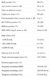
ResultsA total of 140 HBsAg-positive renal transplant patients were followed up at the Hepatitis outpatient clinic of Federal University of Sao Paulo. Ninety-nine (71%) were male, the mean age was 46±10 years (range: 17–74). The patients were included in different time points after renal transplant with a mean time of post-transplant follow-up of 8±5 years. The general characteristics of the patients are shown in Table 1.
General characteristics of the HBsAg-positive renal transplant patients (n=140).
| Male gender (%) | 99 (71) |
| Age (years), mean±SD | 46±10 |
| Time on dialysis (years) | 5.2±3.8 |
| Cadaveric donor (%) | 68 (49) |
| Post-transplant time (years), mean±SD | 8±5 |
| HCV-RNA positive (%) | 28 (20) |
| HBeAg positive (%) | 70 (50) |
| HBV-DNA (log)a, mean±SD | 6.64±2.09 |
| HBV-DNA (%)a | |
| <200IU/mL | 10 (9.5) |
| 200–2000IU/mL | 8 (7.6) |
| ≥2000IU/mL | 87 (83) |
| Fibrosis (F3–4) (%)b | 21 (23.1) |
| Triple immunosuppression (%) | 107 (76%) |
| Immunosuppression (%) | |
| Regimen including MMF | 42/140 (32) |
| Regimen including AZA | 89/140 (63.6) |
| Regimen including CSA | 87/140 (62) |
MMF, mycophenolate mofetil; AZA, azathioprine; CSA, cyclosporine A.
During follow-up, 25% (35/140) of the patients presented elevated ALT, characterizing biochemical exacerbation. This event was observed within a mean period of 3.4±3 years after kidney transplantation (median of two years).
Among the patients presenting exacerbation, viral replication (HBV-DNA and/or HBeAg and/or HBcAg in tissue) was observed in all patients in whom this variable could be analyzed (n=33). Viral load was determined in 20/35 patients by real-time PCR and the median was 29×106IU/mL in these patients.
A liver biopsy was obtained from 83% (29/35) of the patients with biochemical exacerbation. With respect to fibrosis, 76% (22/29) of the patients had fibrosis stage 0–2 and 24% (7/29) had stage 3–4. Mild necroinflammatory activity was observed in 27.5% (8/29) of the patients, moderate activity in 65.5% (19/29), and intense activity in 7% (2/29).
Clinical and/or laboratory signs of hepatic insufficiency were observed in 17% (6/35) of the patients, encephalopathy in 17% (6/35), ascites in 11.4% (4/35), and significant laboratory abnormalities in 14% (5/35). Three of these patients died as a consequence of liver failure despite the use of lamivudine in two of these cases.
Among the 35 patients with exacerbation, 10/35 were not treated with lamivudine. Spontaneous resolution of the biochemical abnormalities without loss of liver function was observed in 9/10 not treated patients and one patient died with liver failure. According to lamivudine use, in only 9% (3/35) of the patients the drug was used as preemptive/prophylactic therapy. Treatment with lamivudine after onset of exacerbation was administered to 22/35 patients, with clinical response in 18/22. The mean time to ALT normalization was 8.8 months in these patients. When lamivudine was given at the time of exacerbation, no difference in mortality due to hepatic insufficiency was observed between treated and untreated patients (9% vs. 10%; p=0.69).
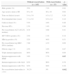
Table 2 lists the clinical and laboratory characteristics associated with biochemical exacerbation. Variables associated with the occurrence of biochemical exacerbation were a less frequent use of mycophenolate mofetil in the immunosuppression regimen and a lower proportion of prophylactic/preemptive administration of lamivudine.
Comparison between patients with and without biochemical exacerbation.
| Without exacerbation (n=105) | With exacerbation (n=35) | p-value | |
|---|---|---|---|
| Male gender (%) | 70% | 71% | 0.92 |
| Age (years), mean±SD | 45±10 | 46±10 | 0.64 |
| Time on dialysis (years) | 5.4±3.9 | 4.5±3.2 | 0.26 |
| Post-transplant time (years) | 7.3±5.0 | 8.9±4.2 | 0.10 |
| Cadaver donor (%) | 51% | 44% | 0.49 |
| Previous RTx | 13% | 14% | 0.99 |
| Pre-exacerbation ALT (×ULN, median) | 0.54 | 0.68 | 0.41 |
| HCV-RNA positive (%) | 23% | 11% | 0.14 |
| HBeAg positive (%) | 52% | 46% | 0.51 |
| Pre-exacerbation log HBV-DNA (median) | 7.68 | 4.53 | 0.09 |
| Fibrosis stage 3–4 | 23% | 24% | 0.87 |
| Triple immunosuppression | 80% | 66% | 0.09 |
| Immunosuppression with MMF | 34% | 17% | 0.05 |
| Immunosuppression with AZA | 63% | 66% | 0.76 |
| Immunosuppression with CSA | 62% | 69% | 0.36 |
| Pre-exacerbation lamivudine (n=41) | 36.2% | 9% | 0.002 |
Bold values indicates level of significance of 0.05 was adopted.
MMF, mycophenolate mofetil; AZA, azathioprine; CSA, cyclosporine; RTx, renal transplantation; ULN, upper limit of normal.
In the logistic regression model only pre-exacerbation lamivudine use was found to be independently associated with biochemical exacerbation (Table 3) showing a protective effect.
DiscussionChronic HBV infection presents an unfavorable course in immunosuppressed patients. Faster progression to hepatic fibrosis and a higher frequency of complications of liver disease have been demonstrated in renal transplant recipients.8 Additionally, cases of reactivation of HBV infection after renal transplantation have been reported, sometimes presenting a fulminant course.17 However, data regarding the frequency and severity of episodes of biochemical exacerbation in renal transplant patients infected with HBV are scarce.
In the present study, biochemical exacerbation was a frequent event in renal transplant patients chronically infected with HBV and was observed in 25% of the patients studied over a follow-up period of 8±5 years. In immunocompetent patients, biochemical exacerbation is frequent and generally related to HBeAg seroconversion.16 Yuen et al. followed up a cohort of 3063 patients and observed biochemical exacerbation in 35% of patients over a mean follow-up period of 29 months.18 In renal transplant patients exacerbation is related to a distinct phenomenon and has a marked negative impact because of the greater severity of these episodes in this specific group of patients.19
In view of the immunosuppression to which they are submitted, renal transplant patients generally present intense viremia,20 with very high levels of HBV-DNA even in HBeAg-negative patients.21 Matos et al.4 demonstrated a viral load higher than 2.000IU/mL in 80% of HBeAg-negative transplant patients with chronic hepatitis B. Most of these cases probably correspond to mutations in the pre-core or core promoter region of HBV since no HBeAg was detected and viral replication was clinically significant.
The occurrence of biochemical exacerbation of HBV infection in renal transplant patients is more related to the immune reconstitution observed after reduction of immunosuppression. Patients receive more intense immunosuppression during the immediate post-transplant period, which promotes a significant increase in viral load associated with a pattern of immunotolerance to HBV. The progressive reduction in the dose of the immunosuppressive agents over time leads to improvement in the immune status and consequent greater hepatocellular damage due to the loss of immunotolerance to the virus.7 In addition, exacerbation might be mediated by other mechanisms, such as hepatotoxicity of the immunosuppressive drugs. In this respect, azathioprine and cyclosporine have been shown to cause liver injury accompanied by a significant increase in aminotransferases.22
In the present study, viral reactivation was observed within an average 3.4±3 years (median of 2 years) after renal transplantation, in contrast to other studies in which this event usually occurred within the first post-transplant year.9–11,20 This finding suggests that exacerbation may not be such an early event, and may be possibly related to modifications of the immunosuppressive regimen as time goes by. This should be considered in the recommendations regarding the duration of prophylactic antiviral treatment, which should be prolonged during the post-transplant period and should not be restricted to the first 12 or 24 months post-transplant.
In addition to its high frequency, biochemical exacerbation was a serious event and was associated with clinical and laboratory signs of hepatic insufficiency in 17% (6/35) of the cases. Three of these patients died as a consequence of hepatic failure. Another study involving the same type of patients also reported a high rate of liver dysfunction (30%) associated with reactivation of HBV infection.11 In the present study it was not possible to identify the factors related to the severity of reactivation, due to the small number of patients with hepatic failure. Thus, all HBsAg-positive kidney transplant patients should be carefully monitored. Biochemical exacerbation needs to be rapidly recognized and control measures should be readily adopted in view of the high morbidity and mortality related to this event.
In view of the high frequency and severity of the biochemical exacerbation that occur in renal transplant patients infected with HBV, it would be important to determine the associated factors in order to allow for early identification of patients at risk of this event. No demographic, epidemiological or laboratory variables could predict the occurrence biochemical exacerbation. In the univariate analysis, the only variables that were associated with this event were the inclusion of mycophenolate mofetil in the immunosuppression regimen, which was less frequent among patients with exacerbation, and preemptive or prophylactic administration of lamivudine, which was also less frequent among patients with exacerbation.
One possible explanation for the less frequent use of mycophenolate mofetil in the immunosuppression regimen among patients with exacerbation might have been the potent immunosuppressive effect of this drug on the host immune response, which would eventually reduce immunomediated hepatocellular damage by more efficiently preventing immune reconstitution over time. Another possibility is related to the antiviral effect of mycophenolate mofetil in inhibiting HBV replication, which has been demonstrated in vitro.23
However, the most important observation in this study was that lamivudine was effective in the prevention of exacerbation as demonstrated by the significantly higher proportion of patients using this drug in the group without exacerbation when compared to the group with exacerbation (36% vs. 9%; p=0.002). This was the only variable independently associated with exacerbation in this study, supporting the recommendation of preemptive/prophylactic administration of antiviral prophylaxis to all patients with chronic HBV infection receiving a renal transplant.
On the other hand, when lamivudine was initiated at the time of exacerbation, no difference in mortality due to hepatic insufficiency was observed between treated and untreated patients. In the study of Han et al.,11 although lamivudine promoted normalization of ALT levels and suppression of viral replication in all treated cases, it did not prevent the progression of histological injury. These data suggest that lamivudine has poor efficacy as a rescue drug in cases of exacerbation and that treatment should preferentially and ideally be initiated before transplantation since immunosuppression has not yet been instituted and the patient therefore presents lower viral loads. Nevertheless, administration of antiviral drugs as early as possible should be considered even for patients who have not received pre-transplant prophylactic/preemptive therapy.
Finally, so far there are no studies evaluating more potent antiviral drugs with a higher genetic barrier to resistance in this particular subgroup of patients. However, this new generation of drugs will probably become the ideal option to prevent exacerbation of HBV infection in renal transplant patients.
Conflicts of interestThe authors declare no conflicts of interest.
The authors wish to thank Dr. Valéria Pereira Lanzoni, Department of Pathology, Federal University of São Paulo, São Paulo, Brazil and Dr. José Osmar Medina de Abreu Pestana, Division of Nephrology, Federal University of São Paulo and Hospital do Rim e Hipertensão, São Paulo, Brazil.


