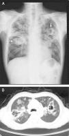
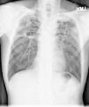
A 59-year-old man with no medical history presented with productive cough and hemoptysis for two months. Low-grade fever, fatigue and body weight loss were noted. Physical examination disclosed crackles in both lungs and cachectic appearance. Blood examination revealed hypoalbuminemia and generalized inflammation with a high C-reactive protein level. Chest radiography and computed tomography showed multiple cavities of various sizes and segmental consolidations in both lungs with upper lobes predominance (Fig. 1A and B). He was admitted under the impression of pulmonary tuberculosis initially. Sputum acid-fast stain and tuberculosis culture did not yield tuberculosis bacilli. Sputum culture yielded Aspergillus fumigatus later and ultrasound-guided fine-needle aspiration of a pulmonary nodule showed hyphae consistent with Aspergillus spp. Chronic pulmonary cavitary aspergillosis was diagnosed. Serial survey did not show evidence of immunodeficiency. After a 6-month course of voriconazole therapy, both symptoms and images remarkably improved (Fig. 2) and body weight increased. Longer duration of antifungal therapy is indicated.
Aspergillosis is an illness caused by aspergillus organisms with various manifestations. Chronic cavitary pulmonary aspergillosis is a multicavitary disease in immunocompetent patients and progresses over time. Previous mycobacterium infections, allergic bronchopulmonary aspergillosis and chronic obstructive pulmonary disease are the most common predisposing conditions.1 Most patients are often suspected of having tuberculosis initially. The common symptoms include cough, shortness of breath, hemoptysis and body weight loss. Radiographic examination usually disclosed multiple cavities of various sizes and ill-defined regions of consolidation, mainly in upper lungs. Diagnosis can be made by classical clinical and radiographic presentations combined with a positive aspergillus serology and/or culture of Aspergillus spp. from the lungs.2 Antifungal agents are the mainstays of therapy. Lifelong therapy is often required.3,4
Conflict of interestAll authors declare to have no conflict of interest.


