Down syndrome (DS) is the most common chromosomal abnormality among live-born infants. In Egypt with 1.6 million births/year, there is estimated risk of 2285 DS births annually.1 Children with DS have an increased risk of infections, especially respiratory tract infections, which can be of diverse pathogenic origin (e.g. viral, bacterial, fungal or a combination of these).2 This increased susceptibility to infections has been linked to abnormal parameters of the immune system, as DS is the most common recognizable genetic syndrome associated with immune defects.3–5
There are few studies on the prevalence and causative pathogens of infections in DS children. We carried out a prospective observational study (March 2011 to March 2012) to determine the prevalence, and risk factors of infections in DS children attending the outpatient genetic clinic in Mansoura University Children's Hospital (MUCH). Our study included 123 (68.7%) males and 56 (31.3%) females. Their ages ranged from 6 months to 11 years. Of these, 68 (38%) patients (44 males and 24 females) had manifestations of infection of the respiratory tract, urinary tract, eye, blood stream, or skin.
Specimens included 35 sputum, 13 throat swabs, 8 conjunctival swabs, 6 urine specimens, 5 blood samples, and one skin swab. Specimens were inoculated on blood agar with optochin disc (Oxoid, UK), incubated at 37°C aerobically, Chocolate agar (Oxoid, UK) incubated in 5–10% CO2 for Haemophilus influenzae, MacConkey agar (Oxoid, UK) for Gram-negative bacilli. Lowenstein Jensen media (Oxoid, UK) was used to exclude Mycobacterium tuberculosis and Sabouraud dextrose agar (Oxoid, UK) for fungi. Laboratory identification of bacterial and fungal isolates was performed by colony morphology, Gram stained films, and biochemical reactions.
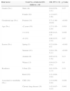
Infections were detected among 68 out of 179 patients (38%). Most infections (60%) occurred during summer. The types of infection were lower respiratory tract infections (LRTIs) (51.5%); upper respiratory tract infections (URTIs) (19.1%); conjunctivitis (11.8%); urinary tract infection (UTI) (8.8%); blood stream infection (7.4%), and skin infection (1.5%). Staphylococcus aureus accounted for 29.4% of isolated pathogens and were most prevalent among skin infections (100%), conjunctivitis (75%) and URTIs (30.8%). Streptococcus pneumoniae came second (14.7%) and were isolated from URTIs (23.1%) and LRTIs (20%). Candida spp. and Escherichia coli were the most frequent pathogens isolated from cases of UTI (33.3%). Of the total bacterial isolates 16.7% were multi-drug resistant (MDR) and they included 4 MRSA, 2 Proteus mirabilis, one Klebsiella pneumoniae, one Pseudomonas aeruginosa and one H. influenzae. Preterm, age less than 2 years or between 2 and 4 years, summer months, congenital heart disease and chronic lung disease were significant risk factors of infection (Table 1).
Risk factors associated with infection among down syndrome children.
| Risk factor | Total No. of infected DS children=68 | OR (95% CI) | p-Value |
|---|---|---|---|
| Gender (No.) | Male (44) | 0.84 (0.58–1.41) | 0.37 |
| Female (24) | 1.12 (0.87–1.46) | ||
| Gestational age (No.) | Preterm (14) | 5.71 (1.96–16.65) | <0.001 |
| Age (No.) | <2 years (30) | 1.57 (1.05–2.28) | 0.019 |
| 2–4 (24) | 0.66 (0.43–1.0) | 0.036 | |
| >4 (14) | 0.96 (0.55–1.52) | 0.87 | |
| Season (No.) | Spring (5) | 0.22 (0.08–0.5) | <0.001 |
| Summer (41) | 2.94 (1.99–4.21) | <0.001 | |
| Autumn (9) | 0.65 (0.32–1.16) | 0.13 | |
| Winter (13) | 0.94 (0.53–1.51) | 0.8 | |
| Residence | Urban (29) | 0.81 (0.56–1.13) | 0.19 |
| Rural (39) | 1.21 (0.88–1.62) | ||
| Associated co-morbidity (No.) | CHD (36) | 1.43 (0.99–2.02) | 0.036 |
| Chronic lung disease (19) | 1.82 (0.97–3.43) | 0.041 |
CHD, congenital heart disease.
In conclusion, respiratory tract infections represent the commonest infection form in DS children in our locality. Also, the emergence of MDR adds additional burden to the problem. Finally, the patterns of infections are seasonally changeable. In line with our findings, we recommend wise use of antibiotics to limit the emergence of MDR bacteria in this group of patients. Furthermore, pneumococcal vaccine should be added to the compulsory vaccination schedule given to DS children in Egypt.
Conflict of interestThe authors declare no conflicts of interest.


