


Nocardiosis is a rare bacterial infection caused by Nocardia spp. However, an increasing incidence has been described whereby data about epidemiology and prognosis are essential.
MethodsA retrospective descriptive study was conducted among patients with positive Nocardia spp. culture, from January 2019 to January 2023, at a Terciary Hospital in Portugal.
ResultsNocardiosis was considered in 18 cases with a median age of 63.8-years-old. At least one immunosuppressive cause was identified in 70% of patients. Five patients had Disseminated Nocardiosis (DN). The lung was the most common site of clinical disease (77.8%) and Nocardia was most commonly identified in respiratory tract samples. The most frequently isolated species were Nocardia nova/africana (n = 7) followed by Nocardia cyriacigeorgica (n = 3) and Nocardia pseudobrasiliensis (n = 3). The majority of the patients (94.4%) received antibiotic therapy, of whom as many as 55.6% were treated with monotherapy. The most frequently prescribed antibiotic was trimethoprim-sulfamethoxazole. Selected antimicrobial agents were generally effective, with linezolid and cotrimoxazole (100% Susceptibility [S]) and amikacin (94% S) having the most activity against Nocardia species. The median (IQR) duration of treatment was 24.2 (1‒51.4) weeks for DN; The overall one-year case fatality was 33.3% (n = 6) and was higher in the DN (66.7%). No recurrence was observed.
ConclusionNocardiosis is an emerging infectious disease with a poor prognosis, particularly in DN. This review offers essential epidemiological insights and underscores the importance of gaining a better understanding of the microbiology of nocardiosis. Such knowledge can lead to the optimization of antimicrobial therapy and, when necessary, guide appropriate surgical interventions to prevent unfavorable outcomes.
Nocardiosis is a rare bacterial infection caused by Nocardia spp., a partially acid-fast, aerobic gram-positive bacillus. It belongs to the Actinomyces genus and is ubiquitous in the environment.1
Nocardia spp. are able to cause localized or disseminated disease, usually affecting immunocompromised patients such as Solid Organ Transplant (SOT) or Hematopoietic Stem Cell Transplantation (HSCT) recipients, patients treated with immunotherapies and/or corticosteroids, with neoplastic disease, Human Immunodeficiency Virus (HIV) infection and structural and functional lung diseases (Cystic fibrosis, Chronic Obstructive Pulmonary Disease [COPD]). Although being generally considered to be an opportunistic pathogen, immunocompetent patients comprise up to one-third of all cases.1
Additionally, despite being considered a rare disease, an increasing incidence has been described, possibly due to increasing number of immunocompromised patients, better awareness and optimization of diagnostic methods. Concurrently, mortality rates are also increasing and can be as high as 50%.1
However, due to the rarity of the disease, data on the prognosis and distribution are still scarce and essential. Therefore, we aim to describe the local epidemiology, clinical characteristics, antibiotic susceptibility patterns and outcomes of patients with infection due to Nocardia spp.
Patients and methodsStudy design and population/data collection/definitionsA retrospective descriptive study was conducted among adults (≥ 18-year-old) with positive Nocardia spp. culture, from January 2019 to January 2023, at Centro Hospitalar de Vila Nova de Gaia e Espinho. This study was approved by the Centro Hospitalar de Vila Nova de Gaia e Espinho ethics committee and a waiver of informed consent was obtained.
Participants were identified by cross-referencing institutional databases of the microbiology and infectious diseases departments. Eligible patients were those who had positive Nocardia spp. culture in any type of bacteriological sample, in addition to compatible clinical and radiological findings of active disease. Patients with clinically suspected nocardiosis, not confirmed by culture or staining were excluded from this study. Other exclusion criteria were incomplete clinical information and loss of follow-up and the identification of Nocardia spp. without clinical or radiological evidence of disease.
Patients’ data was retrieved from medical records and included demographics, smoking habits, alcohol consumption, drug use and comorbidities including underlying pulmonary disease, history of predisposing factors and immunological status (such as HIV infection, SOT, hematological malignancy, active solid tumor, auto-immune diseases, and use of systemic immunosuppressive agents). The presence of bacterial, viral, fungal, and nontuberculous mycobacterial co-infection, treatment course and outcomes were also registered. Taking into account that there were individuals with more than one chronic lung disease, all respiratory diagnoses were classified as preexisting lung disease. The main respiratory disease was defined as the disease that had the greatest clinical, functional, and radiological impact on each patient.
Data regarding the infection consisted in the site of infection, the biological sample in which Nocardia spp. was identified, radiologic exams, antibiotic and treatment duration, and the outcome. The main outcome was death during follow-up or until the disease was considered cured. Nocardiosis was defined as a cause of death if the clinical condition deteriorated, and the patient died from the disease or treatment complications.
DefinitionsNocardiosis was considered a definite diagnosis when Nocardia was repeatedly isolated in multiple samples or in one positive isolate from a sterile site in the presence of a compatible clinical syndrome. Additionally, the patients were classified on the basis of infection type as localized or disseminated.
Lung infection associated with loco regional lymph node involvement was considered as Localized Nocardiosis (LN). Disseminated Nocardiosis (DN) was considered when the infection involved at least two noncontiguous sites with or without pulmonary disease. Central nervous system, bacteremia and hematogenous bone and joint infections were always considered DN even in the absence of other infection sites.
Colonization was considered in patients with Nocardia spp. isolation without clinical or radiological findings consistent with the disease, and these patients were excluded from the analysis.
Immunosuppressive therapy was defined as oncological chemotherapy or treatment with other immunosuppressants (such as azathioprine, methotrexate, or mycophenolate mofetil) within 6-weeks prior to a positive culture for Nocardia spp., or Corticosteroid Therapy (CST). High-dose CST was defined as patients taking a dose equivalent to prednisolone ≥1 mg/kg for more than 21-days or ≥ 10 mg/day during >3 months before the development of nocardiosis.
Efficacy of antibiotic treatment was assessed on the basis of improvement in clinical signs and symptoms and on in vitro sensitivity to antibiotics. Antibiotic regimens were also divided into monotherapy or combined therapies.
Microbiological methodsCultures for bacteriological examination of all samples were carried out using the standard procedure for mycobacteriological examination, with the exception of respiratory specimens (bronchial and bronchoalveolar lavage).
Out of the positive cultures suggestive of Nocardia, Gram and Zielh-Neelsen stains were used for presumptive identification. Definitive identification was made through mass spectrometry (Maldi-Tof, BioMérieux, Portugal) and by molecular Nucleic Acid Amplification Tests (NAAT).
Antimicrobial susceptibility testing was performed by gradient strips (ETEST® BioMÉrieux, Portugal).
For Minimum Inhibitory Concentrations (MIC) interpretation (as Sensitive [S] Intermediate [I] or Resistant [R]) we used the guidance provided by Performance Standards for Antimicrobial Susceptibility Test and Clinical and Laboratory Standards Institute (CLSI).
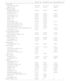
ResultsPatients' characteristicsA total of 25 patients had a suspicion of nocardiosis. Seven patients were excluded, 3 in which Nocardia spp. was not confirmed, 2 due to loss of follow-up and 2 that were considered contamination/colonization. Nocardiosis was considered in 18 cases with a median age on diagnosis of 63.8 years-old (minimum of 34-years-old and a maximum of 84-years-old) and 14 (77.8%) were males. Patients' characteristics are described in Table 1.
Baseline characteristics of patients with documented Nocardia spp. infection.
| Total (n = 18) | Survivors (n = 12) | Non-survivors (n = 6) | |
|---|---|---|---|
| Demographics | |||
| Age, years, mean (SD) | 63.8 (14.8) | 62.1 (15.7) | 67.3 (13.5) |
| Male sex, n (%) | 14 (77.8) | 9 (75.0) | 5 (83.3) |
| Underlying disease | |||
| Diabetes mellitus, n (%) | 9 (50.0) | 4 (33.3) | 5 (83.3) |
| Smoking history, n (%) | |||
| Non-smoker, n (%) | 4 (22.2) | 3 (25.0) | 1 (16.7) |
| Current smoker, n (%) | 6 (33.3) | 4 (33.3) | 2 (33.3) |
| Former smoker, n (%) | 8 (44.4) | 6 (50.0) | 2 (33.3) |
| COPD, n (%) | 6 (33.3) | 4 (33.3) | ‒ |
| Asthma, n (%) | 2 (11.1) | 2 (16.7) | ‒ |
| Bronquiectasis, n (%) | 4 (22.2) | 3 (25.0) | 1 (16.7) |
| Chronic kidney disease, n (%) | 2 (11.1) | 2 (16.7) | ‒ |
| Solid tumor (lung cancer), n (%) | 2 (11.1) | 1 (8.3) | 1 (16.7) |
| Auto-immune disease, n (%) | 3 (16.7) | 3 (25.0) | ‒ |
| HIV infection, CD4+ counts <100 cells/mm3, n (%) | 2 (11.1) | 1 (8.3) | 1 (16.7) |
| Previous OST, n (%) | 1 (5.6) | ‒ | 1 (16.7) |
| Immunosuppresive regimen | |||
| High-dose CSTa, n (%) | 3 (16.7) | 2 (16.7) | 1 (16.7) |
| Immunosuppressive drugsb, n (%) | 4 (22.2) | 3 (25.0) | 1 (16.7) |
| Radiological exams | |||
| Chest CT scan, n (%) | 18 (100) | 12 (100) | 6 (100) |
| Brain imaging, n (%) | 14 (77.8) | 8 (66.7) | 6 (100) |
| FDG-PET/CT, n (%) | 1 (5.6) | ‒ | 1 (16.7) |
| Blood analysis | |||
| Lymphocytopenia, n (%) | 7 (38.9) | 3 (25.0) | 4 (66.7) |
| Site of infection | |||
| Lung, n (%) | 13 (72.2) | 11 (91.7) | 2 (33.3) |
| Central nervous system, n (%) | 1 (5.6) | ‒ | 1 (16.7) |
| Leg abscess, n (%) | 1 (5.6) | ‒ | 1 (16.7) |
| Bone and joint infection, n (%) | 2 (11.1) | ‒ | 2 (33.3) |
| Eye, n (%) | 1 (5.6) | 1 (8.3) | – |
| Type of infection | |||
| Localized, n (%) | 13 (72.2) | 11 (91.7) | 2 (33.3) |
| Disseminated, n (%) | 5 (27.8) | 1 (8.3) | 4 (66.7) |
| Treatment | |||
| Prior prophylaxis, n (%) | ‒ | ‒ | ‒ |
| Monotherapy, n (%) | 9 (50.0) | 8 (66.7) | 1 (16.7) |
| Combination therapy, n (%) | 8 (44.4) | 2 (16.7) | 6 (100.0) |
| Cotrimoxazole, n (%) | 17 (94.4) | 11 (91.7) | 6 (100.0) |
| Imipenem, n (%) | 4 (22.2) | 2 (16.7) | 2 (33.3) |
| Linezolid, n (%) | 3 (16.7) | 1 (8.3) | 2 (33.3) |
| Ciprofloxacin, n (%) | 2 (11.1) | 1 (8.3) | 1 (16.7) |
| Intravenous amikacin, n (%) | 2 (11.1) | ‒ | 2 (33.3) |
| Third generation cephalosporins, n (%) | 1 (5.6) | 1 (8.3) | ‒ |
| Other, n (%) | 3 (16.7) | 3 (25.0) | ‒ |
| No treatment, n (%) | 1 (5.6) | 1 (8.3) | ‒ |
| Overall mortality, n (%) | 6 (33.3) | 2 | 4 |
COPD, Chronic Obstructive Pulmonary Disease; CST, Corticosteroid Therapy; CT, Computed Tomography; FDG-PET/CT, Computed Tomography using 18F-Deoxyfluoroglucose; OST, Organ Solid Transplant.
Underlying diseases were identified in 94.4% of the patients of which Type 2 Diabetes Mellitus (T2DM) was the most common (50.0%), followed by COPD in 33.3% and solid organ malignancy (11.1%). Overall, 72,2% (n = 13) had one or more immunosuppressive conditions including T2DM (n = 9), autoimmune diseases (n = 3), active solid tumors (n = 2) or HIV infection with CD4+ count < 100 cel/uL (n = 2). Among all patients who were immunocompromised, almost one quarter were being treated with high-dose corticosteroid therapy and receiving immunosuppressive agents.
Colonization vs. infectionWe identified two cases of respiratory colonization with N. cyriacigeorgica and Nocardia spp. in two patients with lung cancer. One strain of N. cyriacigeorgica was isolated from the bronchial aspirate of a 65-year-old woman undergoing bronchofibroscopy without any other comorbidities. The Nocardia spp. strain was isolated from a sputum examination. None of these patients received antibiotic treatment.
Among patients with nocardiosis, LN occurred in 13 cases and DN in 5 cases. The lung was the most common site of clinical disease (77.8%). Almost one quarter of these patients had concurrent infections. All patients with a disseminated disease had lung involvement.
At diagnosis, brain imaging workup (cerebral tomography scan or magnetic resonance imaging) was performed in 77.8% (n = 14) of patients. In DN group, 100% of patients had brain imaging, and the lungs and brain were the most frequently involved organs.
Nocardia isolatesNocardia was most commonly identified in respiratory tract samples (83.3%). The most frequently isolated species were Nocardia nova/africana (n = 7) followed by Nocardia cyriacigeorgica (n = 3) and Nocardia pseudobrasiliensis (n = 3). It was not possible to characterize Nocardia subspecies in 2 cases. The location and Nocardia species according to comorbidities of all patients are presented in Table 2.
Patients’ characteristics according to comorbidities, localization of infection, Nocardia species, induction therapy and outcome.
BAL, Bronchoalveolar Lavage; CKD, Chronic Kidney Disease; COPD, Chronic Obstructive Pulmonary Ddisease; CST, Corticosteroid Therapy; DM, Diabetes Mellitus; HIV, Human Immunodeficiency Virus; IS, Immunosuppressive; MFM, Mycophenolate Mofetil; MTX, Methotrexate; OST, Organ Solid Transplant; Cotrimoxazole, Trimethoprim-sulfamethoxazole.
When we specifically analyzed the disseminated group, Nocardia pseudobrasiliensis was the most isolated species (60% of cases) and it was only identified in this group (Table 2).
TreatmentThe majority of the patients (94.4%) received antibiotic therapy, out of whom as many as 55.6% of patients were treated with monotherapy. One patient refused treatment. Therapeutic regimens are shown in Table 1.
The median (IQR) duration of treatment was 34 (6.4‒102) and 24.2 (1‒51.4) weeks for LN and DN, respectively; only one patient with DN received antibiotics for more than 6-months, as the remaining patients died. The disseminated group received mostly combination therapy compared to patients with LN. The most frequently prescribed antibiotics were trimethoprim-sulfamethoxazole (Cotrimoxazole) in 94.4% (n = 17), imipenem (n = 4, 22.2%), linezolid (n = 3, 16.7%) and ciprofloxacin (n = 3, 16.7%).
Selected antimicrobial agents were generally effective, with linezolid and cotrimoxazole (100% Susceptibility [S]) and amikacin (94% S) having the most activity against Nocardia species. In 4 patients, antibiotic susceptibility was not available. The antimicrobial susceptibilities for the Nocardia species are listed in Table 3.
Activities of antimicrobial agents against the 7 most frequently isolated Nocardia species/complexes.
S, Susceptible; I, Intermediate, R, Resistant; Cotrimoxazole, Trimethoprim-sulfamethoxazole; NT, Not tested.
The overall one-year case fatality was 33.3% (n = 6) and was higher in the DN (66.7%). No recurrence was observed. The characteristics of patient's who died are also shown in Table 1.
All of these patients were considered immunosuppressed ‒ mainly due to T2DM. One patient had HIV infection with CD4+ count < 100 cells/mm3 and the other one was a 53-year-old man who received a lung transplant and was under immunosuppressive therapy with 5 mg of prednisone and mycophenolate mofetil. None of these patients were under high dose CST. However, it is noteworthy that 66.7% of DN group presented lymphocitopenia (absolute lymphocyte count < 1 × 10^3/uL).
One patient refused treatment, did not have symptom improvement but is still alive.
DiscussionNocardiosis is an uncommon but emerging disease in both immunocompetent and immunocompromised patients. It may be caused by exogenous inhalation or by direct invasion by injured skin. Therefore, the lung and skin are the most susceptible organs. Ocular and joint infections are rare but have been described.1
The present study provides important information on the risk factors, epidemiological and microbiological characteristics of this disease over a 4-year period. Early recognition and prompt treatment are essential to improve the outcome in this population.
In our study, males were affected more frequently than females, similar to most of the published reports.2-4 The reason for this distribution could be related to men's distinct lifestyle and agriculture-related professions (which increases exposure to Nocardia), hormonal effects on the virulence or growth of this bacteria.2,5
The incidence of nocardiosis is thought to be increasing due to increased awareness and an increased number of immunocompromised patients, such as the growing number of SOT and HSCT recipients.2,6
Among our patients, COPD was the second most common comorbidity (33.3%). Structural modifications of the bronchial architecture, impaired ciliary motility and epithelial damage all lead to impaired local immune defense which may facilitate the presence of Nocardia species.2 Inhaled corticosteroids used for COPD treatment may also increase the risk of Nocardiosis as do shorter courses of systemic corticosteroids during acute exacerbations.7-9
However, it is believed that Nocardia may also colonize the respiratory tract. Therefore, the isolation of Nocardia in sputum should be interpreted carefully. The challenge lies in trying not to routinely initiate antibiotics, but to combine clinical signs, symptoms and chest CT findings to guide this decision.6,10
In agreement with the literature, we also found chronic use of CST to be common in patients with nocardiosis.11,12 However, other factors causing impaired cell-mediated immunity may also contribute to nocardiosis. T-cell mediated immunity is the main protective immune response to nocardiosis explaining the higher rates of nocardiosis in patients with solid organ and HSCT, Acquired Immunodeficiency Syndrome (AIDS), autoimmune diseases and those with lymphoreticular malignancy, with more than 60% of reported cases being associated with one of these conditions.13-15
Evaluation of CD4+ count is helpful in understanding pathogenesis in cases of nocardiosis without comorbidities. The association of idiopathic CD4 lymphocytopenia and nocardiosis is less studied but was already described.16 Moreover, in a recent retrospective multicenter cohort study who aimed to identify the factors associated with Nocardia spp. dissemination, lymphopenia was an independent risk factor for DN and correlates with a worst prognosis.17 In fact, the lower absolute lymphocyte count could imply a more pronounced immunological disfunction, making it a useful index on the evaluation of disease severity.18
In our study, 5 of the 7 patients presenting with lymphopenia had DN and worse outcomes. Hence, CD4+ count should be determined in patients with nocardiosis to evaluate the possible presence of idiopathic CD4+ lymphocytopenia, predicting disease dissemination, a poorer prognosis, and the need for further prophylaxis.
With regard to the site of involvement, pulmonary nocardiosis was the most common type of clinical infection, which is consistent others reports.19-22 However, contrarily to previous reports in which Nocardia farcinica and Nocardia cyriacigeorgica were the most prevalent species worldwide,23 in our report, Nocardia nova/africana was the most frequently isolated Nocardia species. This finding has been described in reports from Australia, but not in studies performed in Europe.24-25
An interesting finding in our study were three cases of disease caused by Nocardia pseudobrasisilensis, which is a rare but emerging species that has been reported in association with more invasive and disseminated disease and higher mortality rates. Moreover, this species is thought to be associated with a less favorable antibiotic susceptibility pattern, which was also described in our analysis.26-28 Although some authors recommend ciprofloxacin as the antibiotic of choice in patients suspected of nocardiosis by this species, we found an isolate resistant to ciprofloxacin, which lead to the inadequate empiric treatment of this patient, and possibly contributed to a worse outcome.29 It is noteworthy that DN resulted in substantial mortality, as 80% of deaths were observed in this group, with Nocardia pseudobrasilliensis being responsible for 40% of deaths in DN.
Antimicrobial susceptibility testing is currently recommended for all Nocardia spp. isolates before initiating antimicrobials, as susceptibilities are often difficult to predict, and patients may not tolerate first line treatments.30-32
Cotrimoxazole, imipenem, linezolid and amikacin, which are recommended as initial treatment for nocardiosis, were the most frequently used antibiotics in our study and the ones with most favorable susceptibility patterns.23,32 Cotrimoxazole is considered the first-line therapy for nocardiosis as most studies have shown high susceptibility rates to this drug, although increasing numbers of cotrimoxazole-resistant Nocardia are being reported.4,33,34
We found high resistance rates to imipenem, ciprofloxacin and amoxicillin-clavulanate, when compared to some of the previous studies, although ceftriaxone resistance was lower than in other reports.35-39
Although we do not use NAAT for the diagnosis of nocardiosis, it may be used in some institutions, and without a culturable specimen, treatment of nocardiosis may be difficult.
Even though most clinical Nocardia isolates are susceptible to cotrimoxazole, linezolid and amikacin, prolonged treatments with these drugs may be challenging. Furthermore, many of the susceptible antibiotics do not have oral formulations, which may lead to increased length of stays in the hospital and decreased quality-of-life.40-43
In our study, treatment-related severe adverse events leading to drug discontinuation occurred in 27.8% of the patients, with acute kidney injury, vomiting and anemia being the most common. All these patients had age >65-years-old. These considerations reveal the difficulty and challenge to manage this condition particularly in elderly patients and in the ambulatory setting. Moreover, they alert to the difficulty of acceptance in home care programs and therefore could lead to prolonged hospital admission – none of our patients could be integrated in these programs.
Strengths and limitationsThis study has some limitations. One limitation is that it pertains to the small sample size, as Nocardia is a rarely encountered opportunistic pathogen and cases of nocardiosis are infrequent.
In addition, due to heterogenicity in patient population and risk factors, it is difficult to extrapolate the findings to general population and hence, this study could not reflect the overall clinical characteristics of different Nocardia species worldwide.
ConclusionNocardiosis is a rare and challenging infectious disease, with a miscellaneous clinical spectrum. The diagnosis is frequently difficult to establish, from suspicion to bacteriological documentation and long-term antimicrobial therapy is often required. This review, the largest Portuguese series so far, demonstrated not only that nocardiosis is responsible for morbidity in Portuguese patients but and also the most frequent and susceptibility patterns of Nocardia species.
We also reported the antimicrobial susceptibility patterns of the first-line and second-line drugs used for this infection.
These findings suggest the need to understand the microbiology of nocardiosis better so that antimicrobial therapy can be optimized, and appropriate surgical intervention be performed, if necessary, to prevent unfavorable outcomes.
Authors’ contributionsAll authors contributed to the conception of the Review.
BB: Data analysis, collection and interpretation, writing, review, and approval of the manuscript. DC, JF, CF, SS, CA, TT, AS, GA and LM: reviewed the literature and drafted the manuscript.
Data confidentialityThe authors declare having followed the protocols in use at their working center regarding patients’ data publication.
Funding sourcesThis research did not receive any specific grant from funding agencies in the public, commercial, or not-for-profit sectors.
Protection of humans and animalsThe authors declare that the procedures were followed according to the regulations established by the Clinical Re-search and Ethics Committee and to the Helsinki Declaration of the World Medical Association updated in 2013.
