To describe clinical, laboratory, microbiological features, and outcomes of necrotizing fasciitis.
MethodsFrom January 1, 2004 to December 31, 2011, 115 patients (79 males, 36 females) diagnosed with necrotizing fasciitis were admitted to Mackay Memorial Hospital in Taitung. Demographic data, clinical features, location of infection, type of comorbidities, microbiology and laboratory results, and outcomes of patients were retrospectively analyzed.
ResultsAmong 115 cases, 91 survived (79.1%) and 24 died (20.9%). There were 67 males (73.6%) and 24 females (26.4%) with a median age of 54 years (inter-quartile ranges, 44.0–68.0 years) in the survival group; and 12 males (50%) and 12 females (50%) with a median age of 61 years (inter-quartile ranges, 55.5–71.5 years) in the non-surviving group. The most common symptoms were local swelling/erythema, fever, pain/tenderness in 92 (80%), 87 (76%) and 84 (73%) patients, respectively. The most common comorbidies were liver cirrhosis in 54 patients (47%) and diabetes mellitus in 45 patients (39%). A single organism was identified in 70 patients (61%), multiple pathogens were isolated in 20 patients (17%), and no microorganism was identified in 30 patients (26%). The significant risk factors were gender, hospital length of stay, and albumin level.
DiscussionNecrotizing fasciitis, although not common, can cause notable rates of morbidity and mortality. It is important to have a high index of suspicion and increase awareness in view of the paucity of specific cutaneous findings early in the course of the disease. Prompt diagnosis and early operative debridement with adequate antibiotics are vital.
Necrotizing fasciitis is a rapidly progressive infectious disease that primarily involves the fascia and subcutaneous tissue. It is an uncommon but life threatening infection. It can affect all parts of body and the lower extremities are the most common sites of infection.1–3 The predisposing conditions are diabetes mellitus, liver cirrhosis, alcoholism, hypertension, chronic renal insufficiency, and malignancy. Prompt diagnosis and early treatment with adequate antibiotic with or without surgical intervention are vital because of high mortality. We herein describe clinical, laboratory, microbiological features, and outcomes of 115 patients diagnosed with necrotizing fasciitis during a consecutive eight-year period and review the relevant literature.
Patients and methodsWe retrospectively reviewed all necrotizing fasciitis cases at Mackay Memorial Hospital, Taitung from January 1, 2004 to December 31, 2011. Demographic data, clinical features, site of infection, type of comorbidities, microbiological and laboratory findings and outcomes were analyzed. The severity of liver cirrhosis was classified according to the Child–Pugh score. Diagnosis was made by operation and based on lack of resistance to blunt dissection of the normally adherent fascia, presence of necrotic fascia, and purulent discharge with a foul fish-water odor. Histopathological findings of surgical specimens typically show neutrophils and bacterial clumps infiltration between collagen bundles with focal necrosis were used to confirm the diagnosis when available. Blood and pus cultures were obtained at the time of first operative debridement. The number of operative debridement, the need for amputation, the duration of hospitalization, and in-hospital mortality rate were also documented.
The continuous variables, presented as medians and inter-quartile ranges (IQR, the range between the 25th and 75th percentile) due to the small sample size, were compared between surviving and non-surviving groups by the Mann–Whitney U test. Likewise the categorical variables were expressed by count and percentage and compared using the Yate's continuity correction or Fisher's exact test. To investigate the independent factors associated with death, simple and multiple logistic regression models were performed. All significant factors on univariate analyses were considered for the initial multivariate models. The final multiple logistic regression model was determined using the backward selection technique, wherein variables that did not improve model fit at p<0.1 were discarded; however, the potential confounders such as age and gender were always forced in all multivariate models for adjustment. Moreover, multicollinearity was also evaluated by variance inflationary factor (VIF). Variables with VIF>5 were then considered to have multicollinearity with other covariates and would be excluded from the multivariate analyses. The statistical analyses were performed with SAS software version 9.2 (SAS Institute Inc., Cary, NC). A two-sided p-value<0.05 was considered as statistically significant.
ResultsClinical findingsOut of 115 cases of necrotizing fasciitis enrolled 91 survived (79.1%) and 24 died (20.9%). There were 67 males (73.6%) and 24 females (26.4%) with a median age of 54 years (IQR, 44.0–68.0 years) in the surviving group; and 12 males (50%) and 12 females (50%) with a median age of 61years (IQR, 55.5–71.5 years) in the non-surviving group, respectively. Table 1 summarizes the clinical features of patients. The most common comorbidity was liver cirrhosis in 54 patients (47%) and diabetes mellitus in 45 patients (39%). Among the 54 patients with liver cirrhosis, 33 patients were chronic alcohol abusers, nine had chronic hepatitis B and 16 had chronic hepatitis C. Eight patients had no comorbidity. Local swelling/erythema, fever, pain/tenderness were the most common clinical features at presentation in 92 (80%), 87 (76%) and 84 (73%), respectively.
Clinical features of the 115 necrotizing fasciitis patients.
| Clinical features | No. of patients (% of total) |
|---|---|
| Local swelling/erythema | 92 (80%) |
| Fever | 87 (76%) |
| Pain/tenderness | 84 (73%) |
| Tachycardia | 43 (37%) |
| Shortness of breath | 32 (28%) |
| Shock | 30 (26%) |
| Bullous lesion | 25 (22%) |
| Consciousness change | 7 (6%) |
| Crepitus | 7 (6%) |
The infection involved the head and neck in four cases (3%), the upper limb in 15 cases (13%), the trunk in 15 cases (13%), the lower limb in 70 cases (61%), bilateral lower limb in four cases (3%) and the perineum and scrotum in 11 cases (10%), as shown in Table 2.
Laboratory findingsAn initial blood count revealed leukocytosis (total white count >12×103/μL) in 60 of the 115 patients (52%), leucopenia (total white count <4×10³/μL) in nine of 115 patients (8%) and thrombocytopenia (platelet count >150×103/μL) in 46 of the 115 patients (40%). Hemoglobin <10mg/dL is observed in 42 of the 115 patients (37%). Prothrombin and activated partial thromboplastin time (>12s and >36s, respectively) were prolonged in 64 (56%) and 44 (38%) of the 115 patients respectively. Acute renal failure was diagnosed in 26 (23%) of the 115 patients but serum sodium and potassium remained normal in most cases. In 74 cases (64%), serum albumin level was below 3g/dL, of whom 18 (16%) were Child–Pugh class C.
Microbiological findingsIsolated microorganisms are summarized in Table 3. A single organism was identified in 70 patients (61%) and multiple pathogens were isolated in 20 patients (17%) and no organism was identified in 30 patients (26%). The most common Gram positive bacteria were group A Streptococcus, followed by methicillin-resistant Staphylococcus aureus (MRSA) and methicillin-sensitive Staphylococcus aureus (MSSA). Escherichia coli was isolated in 11 patients and it was the most common Gram negative bacteria. Aeromonas hydrophila was isolated in seven patients and five patients (71%) died. Vibrio vulnificus was identified in two patients and both have expired. Blood culture was positive in 33 patients (29%): seven among group A Streptococcus and in five cases of Aeromonas hydrophila.
Microorgansims isolated in patients with necrotizing fasciitis.
| Microorganisms | Total (n=115) | Survival (n=91) | Nonsurvival (n=24) | Mortality rate of pathogen (%) |
|---|---|---|---|---|
| Gram positive | 60 (52.2%) | 49 (53.8%) | 11 (45.8%) | 18.3% |
| MRSA | 13 (11.3%) | 10 (11%) | 3 (12.5%) | 23% |
| MSSA | 12 (10.4%) | 11 (12.1%) | 1 (4.2%) | 8.3% |
| CoNS | 1 (0.9%) | 1 (1.1%) | 0 (0%) | 0% |
| Group A Streptococcus | 14 (12.1%) | 9 (9.9%) | 5 (20.8%) | 35.7% |
| Group B Streptococcus | 1 (0.9%) | 1 (1.1%) | 0 (0%) | 0% |
| Group D Streptococcus | 2 (1.7%) | 1 (1.1%) | 1 (4.2%) | 50% |
| Non-group ABD Streptococcus | 6 (5.2%) | 6 (6.6%) | 0 (0%) | 0% |
| α-Hemolysis Streptococcus | 4 (3.5%) | 3 (3.3%) | 1 (4.2%) | 25% |
| Enterococcus | 7 (6.1%) | 7 (7.7%) | 0 (0%) | 0% |
| Gram negative | 57 (49.6%) | 42 (46.2%) | 12 (50%) | 21.1% |
| Escherichia coli | 11 (9.6%) | 10 (11%) | 1 (4.2%) | 9.1% |
| Klebsiella pneumonia | 7 (6.1%) | 5 (5.5%) | 2 (8.3%) | 28.6% |
| Enterobacter species | 4 (3.5%) | 4 (4.4%) | 0 (0%) | 0% |
| Serratia marcescens | 3 (2.6%) | 3 (3.3%) | 0 (0%) | 0% |
| Citrobacter freundii | 3 (2.6%) | 2 (2.2%) | 1 (4.2%) | 33.3% |
| Proteus mirabilis | 7 (6.1%) | 7 (7.7%) | 0 (0%) | 0% |
| Aeromonas hydrophila | 7 (6.1%) | 2 (2.2%) | 5 (20.8%) | 71.4% |
| Vibrio vulnificus | 2 (1.7%) | 0 (0%) | 2 (8.3%) | 100% |
| Morganella | 1 (0.9%) | 1 (1.1%) | 0 (0%) | 0% |
| Pseudomonas aeroginosa | 5 (4.3%) | 4 (4.4%) | 1 (4.2%) | 20% |
| Salmonella group B | 1 (0.9%) | 1 (1.1%) | 0 (0%) | 0% |
| Shewanella putrefaciens | 1 (0.9%) | 1 (1.1%) | 0 (0%) | 0% |
| Acinetobacter species | 2 (1.7%) | 2 (2.2%) | 0 (0%) | 0% |
| Anaerobes | 3 (2.6%) | 2 (2.2%) | 1 (4.2%) | 33.3% |
| Provetella dentiola | 1 (0.9%) | 1 (1.1%) | 0 (0%) | 0% |
| Bacteroides fragilis | 2 (1.7%) | 1 (1.1%) | 1 (4.2%) | 50% |
MRSA, methicillin-resistant Staphylococcus aureus; MSSA, methicillin-sensitive Staphylococcus aureus; CoNS, coagulase negative Staphylococcus.
Surgical debridement and amputation were performed in 102 patients and 8 patients, respectively. Five patients were not operated on. Two patients were too critical to be operated at the time of visit to the emergency department and the family refused operation in three patients due to old age and multiple comorbidities. All patients of Aeromonas hydrophila and Vibrio vulnificus infection received surgical intervention immediately but five of seven Aeromonas hydrophila patients and all Vibrio vulnificus patients died. One, two and three and above three surgical debridement were performed in 31, 33, 35 patients respectively. The mean hospitalization time was 24.5 days (SD 16.29 days).
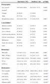
Clinical outcome and factors predictive of deathOf the 115 patients, 24 (20.9%) died and 91 (79.1%) survived. Baseline comparisons between surviving and non-surviving patients with necrotizing fasciitis are shown in Table 4. There were significant differences in age, gender, hospital days, peptic ulcer disease, hemoglobin, platelet, prothrombin time, activated partial thromboplastin time, and all biochemistry tests. The non-surviving group was older, had higher prothrombin time, activated partial thromboplastin time, and glucose, creatinine, aspartate aminotransferase and alanine aminotransferase serum levels (all p≤0.046); additionally, this group had shorter length of hospital stay, and lower hemoglobin, platelet and albumin levels (all p<0.001). The percentage of peptic ulcer disease was higher in the non-surviving group (58.3%) than in the survival group (30.8%, p=0.024).
Summary of baseline characteristics by outcome.
| Survival (n=91) | Death (n=24) | p-Value | |
|---|---|---|---|
| Demographics | |||
| Age (years)a | 54.0 (44.0, 68.0) | 61.0 (55.5, 71.5) | 0.046* |
| Genderb | 0.049* | ||
| Female | 24 (26.4) | 12 (50.0) | |
| Male | 67 (73.6) | 12 (50.0) | |
| Hospital days (days) | 23.0 (15.0, 35.0) | 11.5 (4.0, 25.5) | 0.001* |
| Comorbiditiesb | |||
| Diabetes mellitus | 31 (34.1) | 14 (58.3) | 0.053 |
| Hypertension | 25 (27.5) | 8 (33.3) | 0.756 |
| Liver cirrhosis | 42 (46.2) | 12 (50.0) | 0.916 |
| Cardiovascular disease | 20 (22.0) | 9 (37.5) | 0.196 |
| Chronic renal disease | 26 (28.6) | 12 (50.0) | 0.082 |
| Malignancy | 6 (6.6) | 2 (8.3) | 0.672 |
| Peptic ulce disease | 28 (30.8) | 14 (58.3) | 0.024* |
| Gout | 16 (17.6) | 2 (8.3) | 0.356 |
| Hematologya | |||
| HB (g/dL) | 11.9 (9.5, 13.1) | 9.9 (8.6, 10.8) | 0.001* |
| WBC (103/μL) | 13.9 (9.8, 22.2) | 14.2 (9.7, 21.3) | 0.682 |
| PLT (103/μL) | 243.0 (146.0, 332.0) | 78.0 (48.0, 110.0) | <0.001* |
| PT (s) | 12.0 (11.0, 12.9) | 14.5 (12.9, 16.6) | <0.001* |
| APTT (s) | 33.1 (29.8, 36.0) | 49.4 (39.4, 59.4) | <0.001* |
| Biochemistrya | |||
| GLU (mg/dL) | 120.0 (100.0, 174.0) | 280.0 (198.5, 382.0) | <0.001* |
| Cr (mg/dL) | 1.3 (0.9, 2.4) | 3.6 (2.5, 4.4) | <0.001* |
| AST (IU/L) | 35.0 (19.0, 53.0) | 130.5 (38.0, 161.5) | <0.001* |
| ALT (IU/L) | 24.0 (14.0, 36.0) | 98.5 (20.0, 125.5) | <0.001* |
| Albumin (g/dL) | 2.8 (2.3, 3.1) | 1.7 (1.2, 2.0) | <0.001* |
HB, hemoglobin; WBC, white cell count; PLT, platelet; PT, prothrombin time; APTT, activated partial thromboplastin time; GLU, glucose; Cr, creatinine; AST, aspartate aminotransferase; ALT, alanine aminotransferase.
The continuous data were presented as median (IQR), and compared between different groups by Mann–Whitney U test.
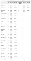
Risk factors associated with death are listed in Table 5. In univariate analysis, gender, hospital days, diabetic mellitus, peptic ulcer disease, hemoglobin, platelet, prothrombin time, activated partial thromboplastin time, glucose, creatinine, aspartate aminotransferase, alanine aminotransferase and albumin were significant predictors of death (all p≤0.034). In multivariate analysis, after adjusting for age and gender, which were forced in the model for controlling for potential confounding effect, the significant risk factors were gender, hospital days and albumin (both p≤0.032). Controlling for age, hospital days and albumin, males had a lower risk of dying than females (OR=0.18, 95% CI: 0.04–0.86, p=0.032); controlling for age, gender and albumin, for every one day increase in hospital stay, death OR decreased by 0.91 (95% CI: 0.85–0.97, p=0.004); controlling for age, gender and hospital stay, for every one g/dL increase in albumin, death OR decreased by 0.05 (95% CI: 0.01–0.18, p<0.001).
Univariate and multivariate logistic regression models for the event of death.
| Univariate | Multivariate | |||||
|---|---|---|---|---|---|---|
| OR | (95% CI) | p-Value | Adjusted OR | (95% CI) | p-Value | |
| Age (years) | 1.03 | (1.00, 1.07) | 0.060 | 1.04 | (0.99, 1.09) | 0.164 |
| Gender (male) | 0.36 | (0.14, 0.90) | 0.030* | 0.18 | (0.04, 0.86) | 0.032* |
| Hospital days (day) | 0.94 | (0.89, 0.98) | 0.003* | 0.91 | (0.85, 0.97) | 0.004* |
| Diabetes mellitus | 2.71 | (1.08, 6.80) | 0.034* | |||
| Hypertension | 1.32 | (0.50, 3.47) | 0.573 | |||
| Liver cirrhosis | 1.17 | (0.47, 2.87) | 0.737 | |||
| CV dis | 2.13 | (0.81, 5.58) | 0.124 | |||
| Chronic renal dis | 2.50 | (1.00, 6.28) | 0.051 | |||
| Malignancy | 1.29 | (0.24, 6.83) | 0.766 | |||
| Peptic ulcer disease | 3.15 | (1.25, 7.95) | 0.015* | |||
| Gout | 0.43 | (0.09, 2.00) | 0.279 | |||
| HB (g/dL) | 0.69 | (0.54, 0.86) | 0.001* | |||
| WBC (103/μL) | 0.98 | (0.93, 1.04) | 0.594 | |||
| PLT (103/μL) | 0.98 | (0.98, 0.99) | <0.001* | |||
| PT (s) | 1.31 | (1.11, 1.55) | 0.002* | |||
| APTT (s) | 1.23 | (1.13, 1.34) | <0.001* | |||
| GLU (mg/dL) | 1.01 | (1.00, 1.01) | <0.001* | |||
| Cr (mg/dL) | 3.24 | (2.00, 5.26) | <0.001* | |||
| AST (IU/L) | 1.03 | (1.02, 1.04) | <0.001* | |||
| ALT (IU/L) | 1.04 | (1.02, 1.06) | <0.001* | |||
| Albumin (g/dL) | 0.06 | (0.02, 0.19) | <0.001* | 0.05 | (0.01, 0.18) | <0.001* |
HB, hemoglobin; WBC, white cell count; PLT, platelet; PT, prothrombin time; APTT, activated partial thromboplastin time; GLU, glucose; Cr, creatinine; AST, aspartate aminotransferase; ALT, alanine aminotransferase; TB, total bilirubin; DB, direct bilirubin.
The comparisons between survival and death in necrotizing fasciitis patients with liver cirrhosis were shown in Table 6. There were significant differences in initial Child–Pugh class between survival and death (p<0.001). The percentage of class C was higher in the non-surviving group (83.3%) than in the survival group (19.0%, p<0.001).
Summary of characteristics by outcome (survival or death) in necrotizing fasciitis patients with liver cirrhosis.
| Survival (n=42) | Death (n=12) | p-Value | |
|---|---|---|---|
| Demographics | |||
| Alcoholism | 25 (59.5) | 8 (66.7) | 0.747 |
| Disease | |||
| Initial Child–Pugh class | <0.001* | ||
| A and B | 34 (81.0) | 2 (16.7) | |
| C | 8 (19.0) | 10 (83.3) | |
| Hepatitis B | 7 (16.7) | 2 (16.7) | 1.000 |
| Hepatitis C | 13 (31.0) | 3 (25.0) | 1.000 |
The categorical variables were expressed by counts and percentages, and compared between different groups by the Fisher's exact test, as appropriate.
The term necrotizing fasciitis was introduced by Wilson in 1952 when he observed a rapid progressive inflammation and necrosis of subcutaneous tissue, superficial fascia, and superficial part of the deep fascia with variable presence of cutaneous gangrene. It has been divided into distinct groups on the basis of microbiological cultures. Type 1 infections are polymicrobial infections that are usually caused by non-group A streptococcus, other aerobic and anaerobic microorganisms. Type 2 infections are usually caused by Streptococcus pyogenes alone or with Staphylococci.4–7
Patients usually present with the triad of pain, swelling, and fever. Tenderness, erythema, and fever are common signs of early necrotizing fasciitis. In our study, local swelling/erythema, fever, pain/tenderness were noted in 92 patients (80%), 87 patients (76%), 84 patients (73%), respectively. It is important to recognize the early stage, which can present with minimal cutaneous manifestations, making prompt diagnosis difficult. Pain out of proportion at the physical examination is the most consistent feature noted at the time of presentation. An apparent cellulitis that does not respond to appropriate antibiotic therapy should raise suspicion of necrotizing fasciitis especially in patients who have an underlying disease. The presence of bullae filled with serous fluid is an important diagnostic clue and should raise the suspicion of this condition. As the infection progresses, the skin characteristically becomes more erythematous, painful and swollen with indistinct borders. The skin develops a violaceous hue, may become necrotic with bullae formation and eventually appears hemorrhagic and gangrenous lesion. But large hemorrhagic bullae, skin necrosis, fluctuance, crepitus and sensory and motor deficits are late signs of necrotizing fasciitis. It is crucial to be alert to these characteristics because the earlier diagnosis of necrotizing fasciitis is made the better outcome and fewer complications will ensue. In our study, 25 patients (22%) had bullae and seven patients (6%) were noted with crepitus.8,9
Necrotizing fasciitis develops not only in the extremities but also in head and neck, trunk, perineum and scrotum. Infections of the head and neck region are associated with high mortality. Mao et al.10 previously reported the poorer survival of patients with thoracic extension (60%) when compared to those without thoracic extension (100%). In our study all patients with head and neck infections have not extended to thorax and no one has died. It might have been due to the alertness of poor outcomes associated with infections of the cranio-cervical region and a more aggressive treatment was implemented in these patients.
Alcohol consumption compromises the integrity of natural barriers to infection in the mouth and decrease saliva flow, resulting in an increased concentration of bacteria which predisposed to cervical necrotizing fasciitis. Although the involvement of extemities is often secondary to trauma, illicit drug use or insect bite, necrotizing fasciitis often develops without any obvious portal of entry in liver cirrhotic patients. Moreover, liver cirrhotic patients usually have chronic edema of the lower limbs, which may predispose them to minor trauma, resulting in an entry port of bacteria. On the other hand, bacteremia may first occur via the intestinal–portal route, because liver cirrhosis weakens the barrier to the passage of bacteria from the intestine to the systemic circulation. Bacteria in the bloodstream may subsequently seed in the edematous soft tissue of lower limbs and then cause infection.11,12 In our study, necrotizing fasciitis in extremities, trunk and perineum occurred in 85 patients (74%), 15 patients (13%), and 11 patients (10%) respectively. It is consistent with other studies.
Some laboratory findings are common in necrotizing fasciitis, but are by no means diagnostic. Anemia, hypoalbuminemia, altered coagulation profile and elevated white cell count were common. In 74 cases (64%), serum albumin level was below 3g/dL, which is probably due to associated malnutrition, compromised liver function due to alcoholism, hepatitis B, C, and the effect of bacterial toxins. Hemoglobin<10mg/dL was noted in 42 of the 115 patients (37%) because the red mass was frequently diminished by thrombosis, echymoses, sequestration by the reticuloendothelial system and hemolysis. Production of red cells by the bone marrow was often depressed by infection and toxemia in these patients. The association of thrombocytopenia, altered coagulative profile and elevated creatinine level with higher mortality found in our study is similar to previously reported articles. It may be due to disseminated intravascular coagulation and toxic shock syndrome. In multivariate analysis, negative prognostic factors for survival were gender, decrease albumin level and decreased length of hospitalization time. It might be due to critical initial presentation with fulminant clinical evolution. Survivors were healthy enough to tolerate further debridement and this increased the length of hospitalization time as found in our series.13–15
Although it is rare, its mortality rate remains high. The etiology is still not fully understood and cannot be identified in many cases. However, it may result from prior history of trauma and certain conditions such as immunosuppression, diabetes mellitus, malignancy, drug abuse and chronic renal disease. Diabetes mellitus is the most common predisposing factor for necrotizing fasciitis and longer hospitalization and higher mortality have been reported. In our study, a higher mortality rate of 58.3% was noted in diabetic patients compared to non-diabetic patients. Hypertensive disease may result in impaired immunity by causing microvascular injury leading to impaired tissue oxygenation and antimicrobial delivery. A mortality rate of 33.3% was found in our hypertensive patients. The percentage of peptic ulcer disease was higher among non-surviving group (58.3%) than in survival group (30.8%).
Group A Streptococcus and S. aureus were the predominant pathogens causing necrotizing fasciitis in the USA and Europe. However, monomicrobial Gram negative aerobic pathogens such as E. coli, A. hydrophila, V. vulnificus were the most frequently isolated microorganisms in Asia. In our series, monomicrobial infections were found in 70 (61%) of 115 patients and polymicrobial infections were found in 20 (17%) of 115 patients. In our study, Vibrio spp. and Aeromonas spp. were not uncommonly detected as the causative organisms, in contrast to other studies. Two patients had Vibrio infection and seven patients had Aeromonas infection. One possible reason for this finding is that Vibrio spp. and Aeromonas spp. are natural inhabitants of seawater, and they are the two common causative pathogens of disseminated bacteremia in patients with liver cirrhosis, and chronic hepatitis. In our study, seven patients had Aeromonas infection and four of them had liver cirrhosis, chronic hepatitis. Two patients had Vibrio infection all had liver cirrhosis and chronic hepatitis. While clinical isolates of A. hydrophila are susceptible to a wide range of antimicrobial agents, they are universally resistant to penicillin, ampicillin, carbenicillin, erythromycin, streptomycin, cefazolin, and clindamycin, and are susceptible to chloramphenicol, ciprofloxacin, co-trimoxazole, aminoglycosides, and third generation cephalosporins. Antibiotic resistance in Aeromonas species poses a potential problem in antibiotic therapy. Intravenous administration of gentamycin or a fluoroquinolone such as ciprofloxacin is recommended for treatment of serious Aeromonas infections, with broad-spectrum penicillins and cefazolin being avoided as first choice agents, particularly for invasive infections. Since in many cases streptococcal or staphylococcal soft tissue infections will be suspected, empirical therapy directed against these organisms most often included penicillin-based antibiotics such as cloxacillin or cefazolin, to which A. hydrophila is intrinsically resistant. The possibility of A. hydrophila infection should be considered when confronted clinically by Gram negative bacilli in purulent exudates and soft tissue swabs. Only then can truly effective antibiotic treatment be provided. Tetracycline and third-generation cephalosporins have been the suggested treatment for Vibrio spp. Once Vibrio infection is suspected, appropriate effective early initiation of antibiotic treatment is important because it significantly improves mortality in Vibrio infection.16–21
Plain X-rays of involved area may show evidence of soft tissue air. In equivocal cases, Computerized Tomography and Magnetic Resonance Imaging are helpful in defining the presence and extent of infection. Although it cannot be overemphasized, these have been used primarily for patients in whom the diagnosis is doubtful. However, the extent of debridement is determined by physical findings at the time of surgery and not by Computerized Tomography findings.
Fournier's gangrene, a necrotizing fasciitis of the perineal, genital and perianal region was first described by Baurienne in 1764. Fournier's gangrene has a high death rate ranging from 15 to 50%, and is an acute urological emergency.22,23 In our series, 11 patients were diagnosed Fournier's gangrene and only three patients survived (27%).
It has been reported to have a mortality rate of 34% (range 6–76%) in necrotizing fasciitis patients and a review involving affection of upper extremities found a mortality rate of 35.7%. Our mortality rate was 20.9%. Death usually occurred as a result of bacterial infection with septic shock, disseminated intravascular coagulation, and/or multiple organ failure. So, early recognition of necrotizing fasciitis followed by appropriate antibiotic therapy with or without surgical intervention is necessary to reduce mortality.24,25
ConclusionDespite being a relatively uncommon infection, the present retrospective study highlights that necrotizing fasciitis can be the cause of notable morbidity and mortality among immunocompromised persons. Aeromonas hydrophila and Vibrio vulnificus infection may be frequently overlooked as the cause of skin and soft tissue infection. The rapid onset of cellulitis in the setting of soft tissue trauma should alert the clinician to the possibility of these two organisms intrinsically resistant to common antibiotics used for cellulitis. Therefore, it is important to have a high index of suspicion and increase awareness at initial presentation. Delayed diagnosis and treatment with adequate antibiotics were crucial for patient survival. Outcomes depend on the promptness of diagnosis, surgical treatment and management of post operative complications.
Conflicts of interestThe authors declare no conflicts of interest.


