
To describe the clinical manifestations and outcome of acute liver failure (ALF) associated with dengue viral infection, a rare but severe complication.
MethodsOne hundred and fifty five consecutive patients with ALF admitted to the national liver centre from 2001 to 2009 were reviewed retrospectively. Eight cases due to dengue infection were identified and their clinical characteristics are described.
ResultsAll patients had severe dengue with one dengue shock syndrome. The median (minimum, maximum) age was 33.5 (17, 47) years with 50% female. The median (minimum, maximum) duration from the onset of fever to development of ALF was 7.5 (5, 13) days and the maximum hepatic encephalopathy (HE) grade were III in five patients and II in three patients. Three patients had systemic inflammatory responses (SIRS) on admission and were in grade III HE. The presence of SIRS on admission was associated with higher grade of HE and its development during the course of hospitalization was associated with worsening HE grade. The hepatitis was characterized by marked elevations in: alanine transaminase [median admission 1140.5u/L (639, 4161); median peak 2487u/L (998, 5181)], serum bilirubin [median admission 29μmol/L (23, 291); median peak 127μmol/L (72, 592)], and prothrombin time [median admission 16.8s (15.3, 26.2); median peak 22s (15.3, 40.7)]. The survival rate with standard medical therapy alone was 100%.
ConclusionsDengue associated ALF manifest about one week after the onset of fever with severe hepatitis and encephalopathy. In our experience, the outcome with standard medical therapy alone is excellent.
Dengue is caused by a small, 50nm diameter single stranded RNA virus of the genus Flavivirus, family Flaviviridae. There are four distinct serotypes called dengue virus type 1 to 4 or DEN-1 to DEN-4. The dengue virus is transmitted to humans through the bites of infected Aedes mosquitoes mainly the female Aedes aegypti.1
The World Health Organization (WHO) estimated that 50 million dengue infections occur annually. In the last 50 years, the incidence has increased by 30 folds with the expansion of geographical distribution to about 100 countries and to the rural areas making it the most rapidly spreading arboviral infection.2 This phenomenon is thought to be contributed by the worldwide expansion of trade and travel which increased the spread of mosquitoes as well as rapid urbanization which increased mosquitoes breeding sites.2
Dengue infection may present as self-limiting acute febrile illness or the more severe forms with dengue shock syndrome, bleeding and other major organ involvements. Gastrointestinal manifestations like hepatitis, acute liver failure, acalculous cholecystitis, acute pancreatitis, acute parotitis and diarrhoea have also been reported.3 Hepatitis is common and can be found in 60–90% of dengue infected patients.4–6 Fortunately, the transaminitis is usually mild to moderate and severe hepatitis with transaminases above ten times the upper limits of normal is less common at 3–11%.5,6
Acute liver failure (ALF) associated with dengue fever has been described, with most reports occurring among children and a few individual case reports in adults.7–14 In the studies involving children, fatal outcomes and a mortality rate of 50% had been reported.15 Although most of the cases reported were from the tropics, one case occurred in a returned international traveller.14
The pathogenesis of dengue associated liver injury is not fully understood but thought to be due to a direct viral effect or from a dysregulated immune response. Histological findings of hepatocytes necrosis at zone two and councilman bodies had been reported.16 Other potential causes of hepatitis in dengue patients are ischaemic or hypoxic liver injury due to circulatory compromise and also drug induced liver injury since medications like acetaminophen or herbal remedies are usually taken for the commonly associated symptoms of fever and body aches.
The aim of this study was to describe in details the clinical characteristics and outcome of a series of adult dengue patients with acute liver failure, a hepatology emergency which is no longer restricted to the tropics due to the increasing travel and trade.
Materials and methodsPatientsFrom 2001 to 2009, all ALF cases above the age of 12 years who were referred to the national liver centre were analyzed. The criteria for diagnosis of ALF were the presence of encephalopathy and coagulopathy (PT>15s) within 26 weeks of the first symptoms without previous underlying liver disease.17
Among the 155 ALF cases, eight were diagnosed to have dengue fever. The diagnosis of dengue fever was based on compatible clinical and laboratory conditions (positive anti-dengue IgM antibody and acute febrile illness with two or more of the following: headache, retro-orbital pain, myalgia, arthralgia, rash, haemorrhagic manisfestations, haemoconcentration, low platelet counts).
The severity of the dengue infection was also classified according to the WHO 2009 classifications: (a) dengue without warning signs, (b) dengue with warning signs or (c) severe dengue which is defined by one or more of the following features: (i) plasma leakage with shock (dengue shock) and/or fluid accumulation with respiratory distress, and/or (ii) severe bleeding, and/or (iii) severe organ impairment.2
Clinical assessmentsConsist of detailed history taking (collateral history and referral notes were used in patients who were encephalopathic) and full physical examinations with attention to pupilary size and response as well as neurological examinations of the lower limbs for tone, reflexes, clonus and plantar responses. Hepatic encephalopathy (HE) was graded according to the West Haven Criteria. The components of systemic inflammatory response (temperatures>38°C or <36°C, white blood count>12,000 or <4000/mm3, pulse rate>90beats/min, respiratory rates>20 breaths/min or PaCO2<4.3kPa) were assessed on admission and at the onset of worsening encephalopathy.
Laboratory and radiological testsDengue serology was done using PANBIO Dengue Immunoglobulin M Capture ELISA. Other laboratory tests carried out were full blood counts, liver function tests, renal profile, coagulation profile, Hepatitis B surface antigen, anti-Hepatitis C antibody and IgM anti-Hepatitis A antibody. Acetaminophen levels were assayed when indicated. Cultures of blood, sputum or tracheal aspirates and urine were carried out once weekly and when clinically indicated. Radiological examinations were transabdominal ultrasonography in all patients and non-contrast CT scan of the brain when indicated.
Ethical approvalSince this study was a case series and retrospective in nature, there are no consent taken from the patients and we received approval from Medical Review and Ethics Committee (MREC) of the Ministry of Health Malaysia.
Data analysisAs this study involved small data sets and the distribution is skewed, the continuous variables were presented in medians with minimum and maximum values while the categorical data were presented in total number and/or percentages.
ResultsDemographics and clinical features at admissionDuring the study period from 2001 to 2009, eight out of 155 adults (5.2%) with ALF were diagnosed to have dengue fever. The median (minimum, maximum) age was 33.5 (17, 47) years with 50% female. All the patients had features of severe dengue with one case in dengue shock according to the WHO 2009 classifications.
Four out of the six patients who could give history, complained of right upper quadrant pain and in one of them, there was clinical and ultrasound evidence of hepatomegaly. One of the remaining two patients who could not provide coherent history also had hepatomegaly. In both cases with hepatomegaly, the liver spans were 16.1cm by ultrasound measurement.
On admission, three patients were in grade III HE while two were in grade II and three did not have HE. All the three patients who were in grade III HE had more than one systemic inflammatory response (SIRS) on admission. The three patients without SIRS criterion were not encephalopathic and the remaining two patients with grade II HE had only one SIRS criterion.
All the patients were tested negative for Hepatitis B surface antigen, anti-Hepatitis C antibody and IgM anti-Hepatitis A antibody. The median (minimum, maximum) acetaminophen levels of the five patients who had taken acetaminophen during the illness were 9.8 (<6.6, 64.4)μmol/L. However these patients did not have the poor prognostic King's College Criteria for acetaminophen overdose when they developed encephalopathy. The other laboratory results at the time of admission are in Table 1.
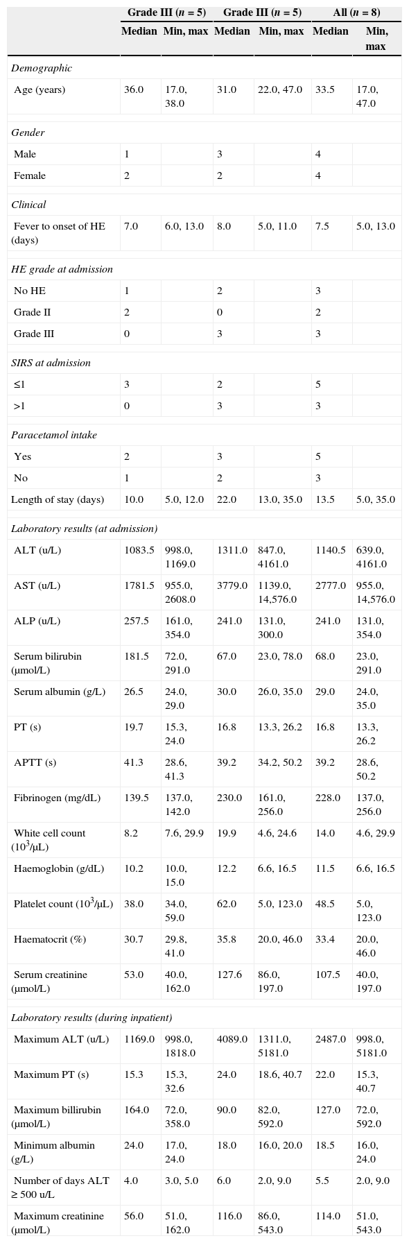
Demographic, clinical and laboratory features of patients at the time of admission and the worst results according to the maximum hepatic encephalopathy grade.
| Grade III (n=5) | Grade III (n=5) | All (n=8) | ||||
|---|---|---|---|---|---|---|
| Median | Min, max | Median | Min, max | Median | Min, max | |
| Demographic | ||||||
| Age (years) | 36.0 | 17.0, 38.0 | 31.0 | 22.0, 47.0 | 33.5 | 17.0, 47.0 |
| Gender | ||||||
| Male | 1 | 3 | 4 | |||
| Female | 2 | 2 | 4 | |||
| Clinical | ||||||
| Fever to onset of HE (days) | 7.0 | 6.0, 13.0 | 8.0 | 5.0, 11.0 | 7.5 | 5.0, 13.0 |
| HE grade at admission | ||||||
| No HE | 1 | 2 | 3 | |||
| Grade II | 2 | 0 | 2 | |||
| Grade III | 0 | 3 | 3 | |||
| SIRS at admission | ||||||
| ≤1 | 3 | 2 | 5 | |||
| >1 | 0 | 3 | 3 | |||
| Paracetamol intake | ||||||
| Yes | 2 | 3 | 5 | |||
| No | 1 | 2 | 3 | |||
| Length of stay (days) | 10.0 | 5.0, 12.0 | 22.0 | 13.0, 35.0 | 13.5 | 5.0, 35.0 |
| Laboratory results (at admission) | ||||||
| ALT (u/L) | 1083.5 | 998.0, 1169.0 | 1311.0 | 847.0, 4161.0 | 1140.5 | 639.0, 4161.0 |
| AST (u/L) | 1781.5 | 955.0, 2608.0 | 3779.0 | 1139.0, 14,576.0 | 2777.0 | 955.0, 14,576.0 |
| ALP (u/L) | 257.5 | 161.0, 354.0 | 241.0 | 131.0, 300.0 | 241.0 | 131.0, 354.0 |
| Serum bilirubin (μmol/L) | 181.5 | 72.0, 291.0 | 67.0 | 23.0, 78.0 | 68.0 | 23.0, 291.0 |
| Serum albumin (g/L) | 26.5 | 24.0, 29.0 | 30.0 | 26.0, 35.0 | 29.0 | 24.0, 35.0 |
| PT (s) | 19.7 | 15.3, 24.0 | 16.8 | 13.3, 26.2 | 16.8 | 13.3, 26.2 |
| APTT (s) | 41.3 | 28.6, 41.3 | 39.2 | 34.2, 50.2 | 39.2 | 28.6, 50.2 |
| Fibrinogen (mg/dL) | 139.5 | 137.0, 142.0 | 230.0 | 161.0, 256.0 | 228.0 | 137.0, 256.0 |
| White cell count (103/μL) | 8.2 | 7.6, 29.9 | 19.9 | 4.6, 24.6 | 14.0 | 4.6, 29.9 |
| Haemoglobin (g/dL) | 10.2 | 10.0, 15.0 | 12.2 | 6.6, 16.5 | 11.5 | 6.6, 16.5 |
| Platelet count (103/μL) | 38.0 | 34.0, 59.0 | 62.0 | 5.0, 123.0 | 48.5 | 5.0, 123.0 |
| Haematocrit (%) | 30.7 | 29.8, 41.0 | 35.8 | 20.0, 46.0 | 33.4 | 20.0, 46.0 |
| Serum creatinine (μmol/L) | 53.0 | 40.0, 162.0 | 127.6 | 86.0, 197.0 | 107.5 | 40.0, 197.0 |
| Laboratory results (during inpatient) | ||||||
| Maximum ALT (u/L) | 1169.0 | 998.0, 1818.0 | 4089.0 | 1311.0, 5181.0 | 2487.0 | 998.0, 5181.0 |
| Maximum PT (s) | 15.3 | 15.3, 32.6 | 24.0 | 18.6, 40.7 | 22.0 | 15.3, 40.7 |
| Maximum billirubin (μmol/L) | 164.0 | 72.0, 358.0 | 90.0 | 82.0, 592.0 | 127.0 | 72.0, 592.0 |
| Minimum albumin (g/L) | 24.0 | 17.0, 24.0 | 18.0 | 16.0, 20.0 | 18.5 | 16.0, 24.0 |
| Number of days ALT≥500u/L | 4.0 | 3.0, 5.0 | 6.0 | 2.0, 9.0 | 5.5 | 2.0, 9.0 |
| Maximum creatinine (μmol/L) | 56.0 | 51.0, 162.0 | 116.0 | 86.0, 543.0 | 114.0 | 51.0, 543.0 |
Min, minimum; Max, maximum; HE, hepatic encephalopathy; SIRS, systemic inflammatory response score; ALT, alanine transaminase; AST, aspartate transaminase; ALP, alkaline phosphatase; PT, prothrombin time, control, 11.2s; APTT, activated partial thromboplastin time, control, 33.9s. Gender, HE grade at admission, SIRS at admission and paracetamol intake were reported in frequency.
One of the three non-encephalopathic patients progressed to grade II HE on day four of admission and two deteriorated to grade III HE on day two and four respectively. All of them had more than one SIRS criterion when the grade of HE worsened, while the two grade II HE patients who did not progress into higher grades of HE, did not develop SIRS.
Therefore the maximum grade of HE was grade III in five patients and grade II in three patients, all occurring within one to four days of admission to our hospital. The median (minimum, maximum) duration from the onset of fever to the development of HE or ALF was 7.5 (5, 13) days. All the five grade III HE patients were electively intubated for cerebral protection. Lower limbs hyperreflexia and sustained clonus were found in three patients and one had bilateral upgoing plantars response. Pupilary abnormality was documented in one patient only. Non-contrast CT scan of the brain was carried out in four of them and the findings were normal.
During the course of the illness, one patient developed ascites and pleural effusion, two patients had ascites alone and another two had pleural effusions alone. One male patient aged 38 with peak values for alanine transaminase (ALT) 1169u/L, aspartate transaminase (AST) 2608u/L, prothrombin time of 38s and grade II HE developed melena. An emergency upper endoscopy revealed altered blood with mucosal sloughing in the stomach.
Six patients (75%) acquired culture proven infections and the sites of positive cultures were respiratory (n=4), blood (n=3) and urine (n=2). All these patients had presence of SIRS. The remaining two patients who never had SIRS, did not have any positive cultures.
The peak values of the liver and renal parameters during the hospitalization and the duration of time with ALT≥500u/L are presented in Table 1.
Clinical managementsAll the patients were nursed with the head of bed elevated at 30° and enteral nutrition support was initiated whenever possible. Fluid managements with crystalloids and colloids and correction of coagulopathy with fresh frozen plasma and platelets were carried out as clinically indicated. All but one patient received N-acetylcysteine infusion at 100–150mg/kg/day for a minimum of five days. Broad spectrum antibiotics were started on all patients and fluconazole was given to six patients. Three patients received mannitol and 3% saline infusions for clinical signs of raised intracranial pressure. Only one of the three patients with renal impairment required renal replacement therapy during the acute illness.
Outcomes and follow-up dataThe median (minimum, maximum) duration of intubation in the five patients with grade III HE was 11 (2, 17) days. All the patients were discharged well after 13.5 (5, 35) days of hospitalization and the seven patients who were followed up had normalization of ALT with a median value of 24u/L (14, 38).
DiscussionIn this series of eight patients, we found that adult dengue patients who developed acute liver failure did so at a median of 7.5 days (5, 13) after the onset of fever with some occurring in the recovery phase of dengue illness. According to the WHO 2009 classifications, all our patients had features of severe dengue infection. These findings are comparable to other case reports which reported ALF at five to 14 days from the onset of fever and associated with severe types of dengue infections.10–13
Interestingly similar to acute liver failures of other aetiologies, we found that the presence of SIRS in dengue related ALF is also associated with higher grade of hepatic encephalopathy, with worsening of encephalopathy and positive microbiology.18
After a period of three to seven days incubation, the course of dengue illness typically starts with an acute febrile phase for two to seven days. This is followed by a critical phase at around the time of defervescence starting from three to seven days of the illness during which there are increased capillary permeability and systemic vascular leak. Those who survived through the 24–48h of clinically significant plasma leakage in the critical phase will enter the recovery phase with rapid improvement in symptoms.2,19 The dengue virus and the virus-encoded NS1 are present in the blood during the acute phase of illness.19 A study by Thomas et al. on two serotypes of the dengue virus, DEN-2 and DEN-4, showed that the dengue viral load fell from day one to six of illness and more severe presentation was associated with higher viral load.20
In our patients, the maximum grade of encephalopathy occurred on either the same day (n=6) or one to two days (n=2) after the ALT peaks. These were at five to 13 days from the onset of fever which may coincide with about one week after the viral surge. Therefore, health care providers who are looking after severe dengue cases with hepatitis should look out for early signs of ALF in the recovery phase of dengue illness at around or shortly after the time of ALT peaks together with the development of SIRS criteria.
Our patients had markedly elevated serum transaminases at values above 10 times the upper limit of normal and the AST levels were higher than ALT as found in the few case reports of dengue associated ALF.7–14 The AST levels are also higher than ALT in the other milder form of dengue hepatitis.4,6,21 The AST had been reported to peak at about seven to eight days after the onset of illness and the ALT lags behind AST in time and magnitude.6
We did not find co-infection with hepatitis A, B or C in our ALF patients. Acute and chronic hepatotrophic viral hepatitis are common in this region, and there are valid concern that concomitant infection will results in more severe liver disease. Two older studies reported that hepatitis B or C co-infection did not affect the level of transaminitis.4,6 However, two recent studies showed significantly higher ALT levels in dengue patients with chronic hepatitis B compared to those without.21,22 Luckily, the studies did not find any detrimental effect from the higher transaminases on the coagulation profile or severity of liver disease.21
Five of our patients developed ascites or pleural effusion or both, these were probably due to plasma leakage in the critical phase of dengue illness, the low oncotic pressure and volume overload. Plasma leakage is a feature of severe dengue fever.2 Transient endothelial dysfunction occurs in dengue illness and it can cause hypoalbuminemia and proteinuria.19,23,24
Our patients received N-acetylcysteine infusion as part of the medical therapy for ALF without adverse effects. Kumarasena et al. reported similar experience in dengue ALF.25 Apart from acting as an antidote to paracetamol poisoning, N-acetylcysteine had been proven to improve systemic haemodynamics and tissue oxygen delivery in acute liver failure.26,27 Subsequently randomized double-blind, placebo controlled study had shown that intravenous N-acetylcysteine improved transplant free survival and well tolerated in non acetaminophen ALF when given in early stage.28
In our series of eight adults with dengue associated acute liver failure, the outcome was good with 100% spontaneous survival from standard medical therapy alone. This is in contrast to studies on children which reported 50–66% mortality.15,29 One of the reasons for better outcome in adults may be due to the generally higher mortality from dengue infection in children.30,31 The host risk factors for severe dengue are young age, female gender, high body mass index and certain host genetic variants. The viral factors were the virus strain, and a secondary dengue infection.19
The limitation of our study is the small number but these cases are from the country's only tertiary liver centre and over a study period of nine years. In Malaysia, the incidence of reported dengue cases between 2001 and 2008 ranged from 72.2 to 180 per 100,000 population, so dengue associated ALF is truly a rare manifestation of dengue fever.32
Since this is a rare condition with global concern, we hope a detailed description of the clinical findings in this case series will assist clinicians working in countries with heavy burden of dengue as well as those practising in temperate regions who may come across an ill returned traveller with ALF. Being aware of the features of dengue associated ALF may help to diagnose the condition early. This is important as we found that dengue associated ALF can occur up to 13 days after the onset of illness.
Conflict of interestAll authors declare to have no conflict of interest.
We thank the Director General of Health of Malaysia for permission to publish this article and the Clinical Research Centre of the Malaysian Ministry of Health for statistical and other support.
