
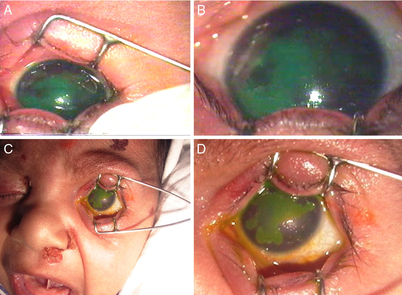
A 14-day-old neonate presented with excessive crying and not opening the eyes since last one week. She was afebrile and had a normal overall general condition. There was a vesicular rash near the left nasolabial fold. On examination under anesthesia, large geographical ulcer involving three-fourths of the cornea with stromal edema was present in both the eyes (Fig. 1). The pupil and iris pattern were normal. Keeping the possibility of primary herpes simplex keratitis, direct immunofluorescent staining of vesicle fluid was done. The test was positive for herpes simplex virus (HSV) 2. Cerebrospinal fluid (CSF) examination was normal including the PCR for HSV. On probing the history, the antenatal period was uneventful, and the mother did not have any history of fever, rash, vaginal discharge or genital lesion. However, maternal IgG was positive for HSV 1 and 2 antibodies. Both topical and systemic acyclovir were prescribed. After three weeks, the skin and corneal lesions had healed.
 Right eye showing a large geographic ulcer (stained). (C and D) Left eye showing a large geographic ulcer (stained). (C) Vesicular rash near left nasolabial fold.")
Many neonatal infections occur because of asymptomatic cervical shedding of virus, usually after a primary episode of HSV infection.1 Diagnosis requires a high index of suspicion because the history of an active infection, primary or secondary, in a mother is often not given, as in the index case. Any vesicular rash in an infant up to eight weeks of age should be cultured and the infant immediately started on antiviral therapy with acyclovir pending culture results. Ocular involvement occurs in 20% of cases of neonatal herpes.1,2 The infant is protected for the first few months of life by circulating maternal antibodies, but this might not be the case always.1 Ocular involvement ranges from mild keratoconjunctivitis to severe retinitis.2 Rarely, bilateral ocular involvement can be seen. The diagnosis is entirely clinical, and prompt therapy is essential because of risk of dissemination.3
Conflicts of interestThe authors declare no conflicts of interest.
