
HEV is one of the most important causes of acute clinical hepatitis in adults with particularly high mortality in pregnant women in developing countries, such as China.1 Although low seroprevalence of HEV infection in pregnant women was reported in Kunming City, Yunnan Province, China in 2011,2 an outbreak of HEV occurred in the city in 2015 affecting both pregnant women and general population. In the present study, the prevalence of HEV was investigated in Kunming city by ELISA and RT-PCR methods. Results indicated that HEV infection epidemic in Kunming city was associated with swine HEV, and pregnant women in their third trimester were more susceptible to HEV infection.
Serum samples of asymptomatic pregnant women (n=274, 90 samples in the first, 92 samples in the second, and 92 samples in the third trimesters of pregnancy) and general population (n=114) were collected for HEV antibodies and HEV RNA detection from January to April 2015.
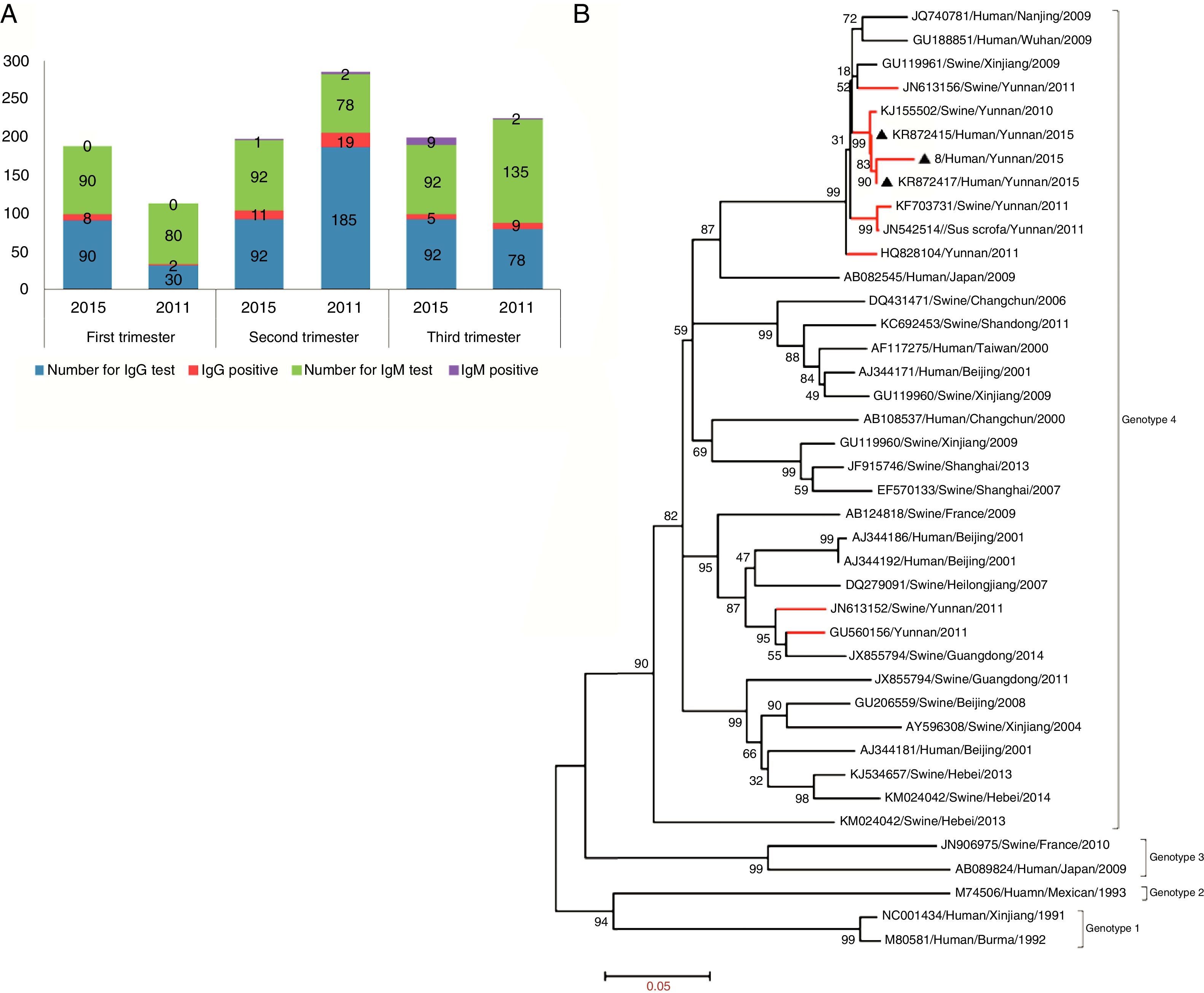
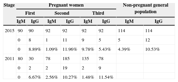
IgM antibody was detected during the acute stage of HEV infection. Among samples of the pregnant women, 10 sera (10/274, 3.65%) were positive to HEV IgM, which was similar to the general population in 2015 (5/114, 4.39%), but higher than that in 2011 in the same city (4/293, 1.37%),2 and Shandong Province, China in 2011–2013 (2.6%).3 Surprisingly, 90% of pregnant women positive for HEV IgM antibody (9/10) were in their third trimester of pregnancy, which was significantly higher than in other stages of pregnancy (Table 1). Furthermore, the activity of AST in HEV IgM positive pregnant women (40–20IU/L) was higher than that in general population (<20IU/L). Results suggested that pregnant women in their third trimester of pregnancy were more susceptible to HEV infection and caused more severe liver lesions, which may partly explain the extremely high mortality rate in the third trimester.
Seroprevalence of HEV in pregnant women in Kunming city.
| Stage | Pregnant women | Non-pregnant general population | ||||||
|---|---|---|---|---|---|---|---|---|
| First | Second | Third | ||||||
| IgM | IgG | IgM | IgG | IgM | IgG | IgM | IgG | |
| 2015 | 90 | 90 | 92 | 92 | 92 | 92 | 114 | 114 |
| 0 | 8 | 1 | 11 | 9 | 5 | 5 | 12 | |
| 0 | 8.89% | 1.09% | 11.96% | 9.78% | 5.43% | 4.39% | 10.53% | |
| 2011 | 80 | 30 | 78 | 185 | 135 | 78 | ||
| 0 | 2 | 2 | 19 | 2 | 9 | |||
| 0 | 6.67% | 2.56% | 10.27% | 1.48% | 11.54% | |||
Among the serum samples, 8.76% (24/274) of pregnant women and 10.53% (12/114) of the general population were positive to HEV IgG antibody, which was similar to the rate found in 2011 in this city (30/293, 10.24%, p>0.05). The positivity rates of anti-HEV IgG antibody in the first, second, and third trimesters of pregnancy were 8.89% (8/90), 11.96% (11/92), and 5.43% (5/92), respectively (Fig. 1A). The highest rate of positive anti-HEV IgG antibody (11/24, 45.83%) was also in the second trimester of pregnancy, similar to that investigated in 2011 in the same city (Fig. 1A).
 Seroprevalence of HEV in pregnant women in Kunming city in 2011 and 2015. (B) Phylogenetic analysis based on nearly full-length (7067bp, exclude 5′ UTR and 3′ poly (A)) nucleotide sequence of isolates in this study and other 37 references of four genotypes of HEV, using the neighbor-joining method. The tree was evaluated using the interior branch test method with Mega 4 software. The scale bar represents nucleotide substitutions per base. GenBank accession number, origin, and host are indicated. The isolates identified in this study are marked with a triangle. Strains isolated from Yunnan province are marked with red color. Isolate 8 was deposited into GenBank database and is still being processed.")
Prevalence of HEV infection in Kunming city and phylogenetic analysis. (A) Seroprevalence of HEV in pregnant women in Kunming city in 2011 and 2015. (B) Phylogenetic analysis based on nearly full-length (7067bp, exclude 5′ UTR and 3′ poly (A)) nucleotide sequence of isolates in this study and other 37 references of four genotypes of HEV, using the neighbor-joining method. The tree was evaluated using the interior branch test method with Mega 4 software. The scale bar represents nucleotide substitutions per base. GenBank accession number, origin, and host are indicated. The isolates identified in this study are marked with a triangle. Strains isolated from Yunnan province are marked with red color. Isolate 8 was deposited into GenBank database and is still being processed.
One third of IgM positive people (5/15) were also positive to HEV RNA. The complete sequences of HEV, two strains isolated from pregnant women, and one strain from non-pregnant people of the general population were obtained by RT-nPCR according to our previous study.4 Sequences were submitted to the GenBank database (KR872417 and KR872415 isolated from pregnant women). Phylogenetic analysis indicated that the three human HEV strains belong to HEV genotype 4, sharing 99.3–99.8% homology with each other and 96.8–99.4% similarity to swine HEV strains isolated from 2009 to 2011 in this city (Fig. 1B). This result indicates that the source of HEV infection in Kunming city in 2015 may originate from the same strain, probably a swine strain.
The high prevalence of HEV infection in Yunnan province may be associated with the traditional raw pork meat consumption and swine-breeder liver in the same house. The third national viral hepatitis prevalence survey of China during 2005–2006 revealed that Yunnan had one of the highest seroprevalence of HEV in China.1 Consuming raw pig livers or raw pork meat have a high potential risk of HEV infection; about 78.9% of the swine in Kunming City tested positive to HEV IgG antibody5 and raw pork meat is consumed frequently as a traditional food during the Spring Festival. Therefore, Hepatitis E should be considered in the differential diagnosis during pregnancy in order to prevent the loss of mother or baby. Infection was highly associated with the prevalence of swine HEV infection in this region, underscoring the zoonotic transmission of HEV.
Conflict of interestThe authors declare no conflicts of interest.
We thank all the patients who actively participated in this study. This study was supported by National Natural Science Foundation of China (31360619), Natural Science Foundation of Yunnan province in China (Grant 2011FZ068, 2013FB032 and 2013FZ142), and China Postdoctoral Science Foundation (2014M562672 and 2015T81138).
